By -1.Bibha Kumari and 2.Ranveer Kumar Sinha
1. First and corresponding author. Mail I.D.- bibhababyvet@rediffmail.com
Subject Matter Specialist (Veterinary Science), K.V.K., Arwal,
B.A.U., Sabour, Bhagalpur (Bihar)
 2. Assistant Professor, Department of Veterinary Medicine
B.V.C., B.A.S.U., Patna (Bihar)

1. Introduction:
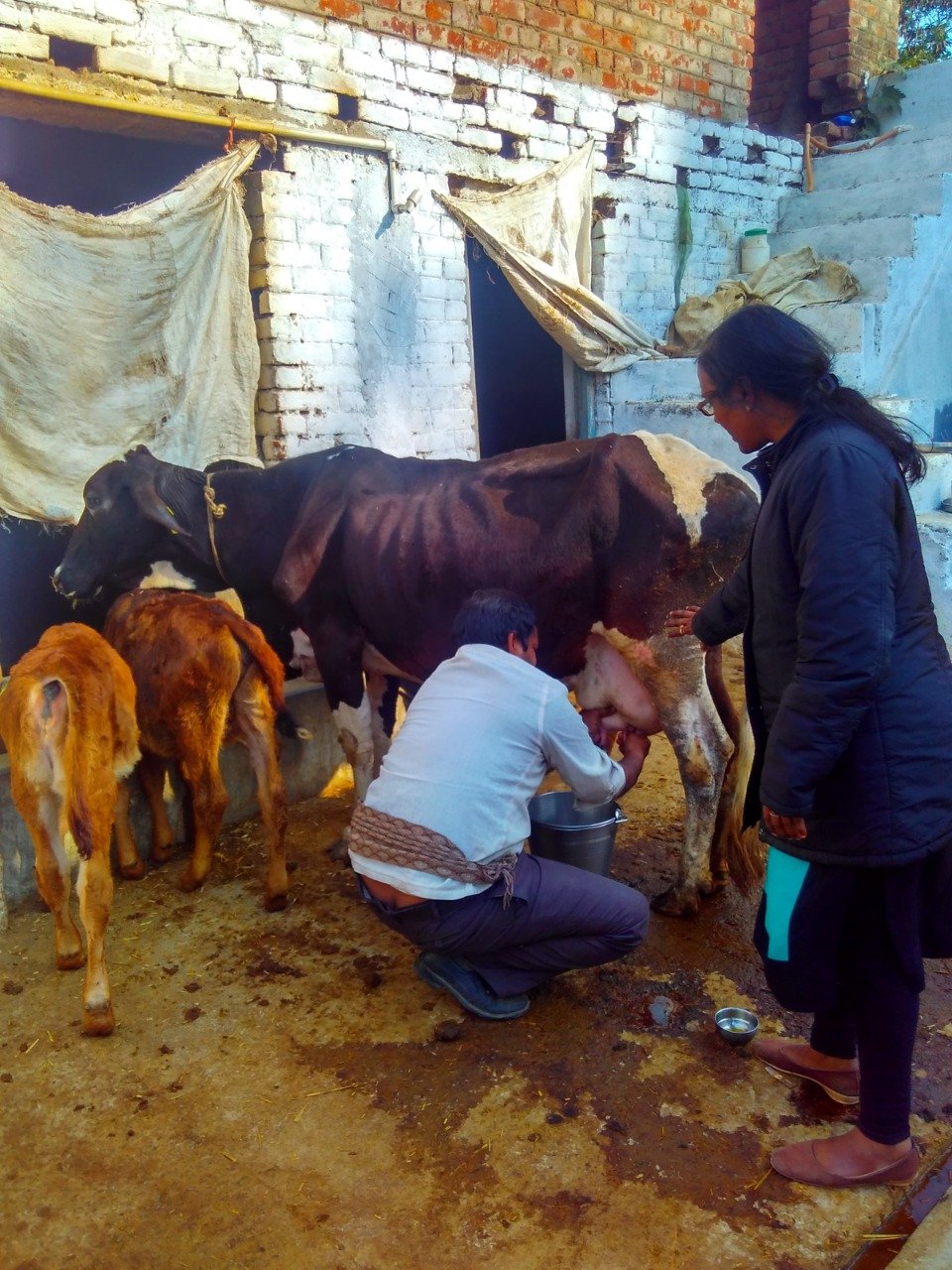
Mastitis is an inflammatory reaction of the udder tissue caused by infection, toxin or trauma of udder. It is one of the most wide spread and common disease characterized as endemic disease affecting dairy herds worldwide (Halasa et. al., 2007). It leads to Physical, chemical and bacteriological changes in milk and pathological changes in glandular tissue of udder. Mastitis is a most challenging disease in high producing dairy cattle in India next to Foot and Mouth disease (Varshney & Mukherjee, 2002). But according to report of Sharma,2003 mastitis placed first position in prevalence of dairy animal disease as more than 90 % high yielding cows suffered with mastitis. The disease leads to accountable economic losses by reduced milk yield (up to 70 %), milk discard after treatment (9%), cost of Veterinary services (7 %) and premature culling (14 %) of animal (Bhikane and kawitkar, 2000). Occurrence of mastitis mostly due to several species of bacteria and also due to fungal, yeast or viral infection. Over use of antibiotics and poor sanitation leads to yeast mastitis (Ganguly, 2018).
A number of factors affecting susceptibility to mastitis are:
• Physiological status of cow.
• Level of milk production.
• Parity of cow.
• Inheritant feature.
• Environmental condition.
2. Etiology
Mastitis is a multi-etiological disease. It can be caused due to different contagious and environmental micro-organisms such as Gram-positive cocci, Gram-negative cocci (mainly coliform group) and other miscellaneous organism like fungus, yeast and virus.
Mastitis can be categorized in two major groups :
• Contagious mastitis:- The causative organisms living on the skin of the teat and inside the udder. They can be transmitted from one cow to another during milking.
• Environmental mastitis:- The causative organism not live on the skin or in the udder but enter the teat canal when the cow comes in contact with contaminated environment. These pathogenic organisms found in faeces, bedding material and feed.
3. Mode of Transmission
• Through teat canal.
• Fly and insects.
• Contaminated bedding materials.
• Contaminated milker’s hands and cloths.
• Contaminated machine cup by affected quarter.
4. Economic Loss
Following factors are responsible for economic losses by dairy man due to mastitis:-
• Reduced milk production.
• Milk discard due to abnormal characteristics and presence of antibiotics.
• Decrease market value of cow due to damage of quarters.
• Cost of Veterinary services and drugs.
• Increased man power consumption to care infected cow.
5. Symptom:

According to symptom mastitis divided into two groups:-
1. Clinical Mastitis :-
It is characterized by visible change in milk, udder or teats. It is further classified as :-
• Per acute mastitis: –
Characterized by painful swelling of udder, fever(105-1060F) shivering, anorexia, depression, cessation of milk secretion and blood stained exudates from teat canal.
• Acute mastitis: –
It is similar to Per acute mastitis but systemic sign like fever, depression are not seen, Udder become swollen and milk secretion changed to curdy yellow material or brown fluid with flakes or clots. Infection may be in one quarter or entire udder.
• Sub acute mastitis: –
There is a variable change in the milk but no Practical changes seen in udder and visible systemic sign. Cultures of milk only show presence of pathogenic bacteria.
• Chronic mastitis: –
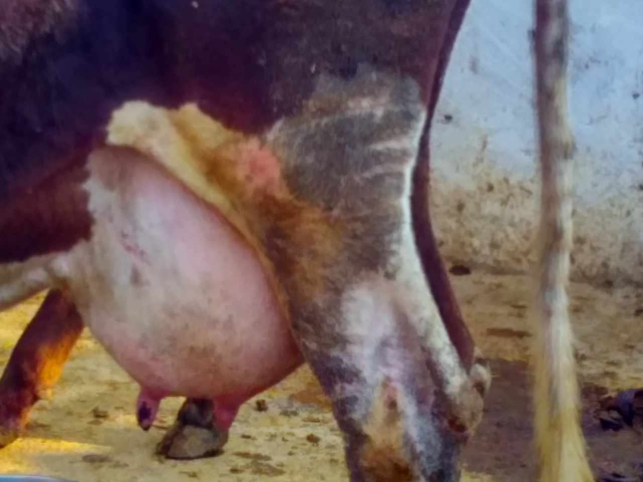
It occurs due to persistent infection of udder. Udder becomes hard due to fibrosis. The quarters may become thickened, firm, nodular and atrophic.
2. Sub-clinical mastitis: –
It is characterized as change in milk composition without any visible change in udder or milk. Sub-clinical mastitis reducing milk production, decrease milk quality and suppress reproductive performance. A high somatic cell count (SCC) is indicative of sub-clinical mastitis.
6. Diagnostic trends
1. Physical examination of the udder.
2. Test for milk abnormalities:-
• Strip cup test
• PH test
• Chloride test
• California mastitis test (CMT)
• Bromothymol blue test (BTB)
• Bromocresol purple test
• White side test
• Hotis test
3. Direct test:-
• Isolation and identification of the organism
• Cultural examinations.
• Biochemical test
• Serological test
• Electrical conductivity test (EC test)
• Somatic cell count (SCC)
7. Treatment
• Remove secretion as much as possible from affected quarter. Sterile test siphon may be used to drain out the milk/secretion. Oxytocin may be used for complete let down of milk and flushing out of inflammatory debris.
• Culture and antibiotic sensitivity test of milk for selection of antibiotics.
• Intramammary (IMM) antibiotic for 3-5 days.
• Systemic antibiotics (IV, IM, SC) for 3-5 days.
• Systemic anti-inflammatory drug for 3-5 days.
• Antihistaminic drug.
• Corticosteroid may be given to check fibrosis.
• Enzyme like Serratopeptidase and Hylase to digest the pus.
• Immunomodulator preparation containing Vit.- E and Se for 4 days.
• Topical application of anti-inflammatory ointment twice a day for 5-7 days.
• Drying off quarters which do not respond to treatment by silver nitrate solution or cupper sulphate solution.
8. Preventive Measures
• Washing the udder and hand of milker with antiseptic before and after milking.
• Infected cow/teat should be milked at last.
• First strip of milk should not allow falling on the floor. It should be stripped in separate container.
• Mastitis milk should be properly disposed. 5% phenol may be added to the infected milk at the time of disposal.
• Dipping of all teats following each milking with Iodophor solution containing 1% available iodine or Hypochlorite solution.
• The animal should not allow lie down immediately after milking for an hour by engaging with feed and fodder.
• Cleaning and disinfecting milking machine after each milking.
• Dry cow therapy to prevent occurrence of mastitis after parturition.
• Newly purchased cows should be kept and milked separately until screened for mastitis(CMT).
• Cow should allow soft bedding following parturition.
• Concrete floor predisposed to mastitis. Bedding should be done with straw, saw dust or sand. Sand is the best, since it has lower bacterial content.
• The non responsive quarter should be permanently dried up.
9. Conclusion
Mastitis is a disease caused by many agents. There is the interaction between management practice and infectious agents in occurrence of mastitis. A combination of preventive measures and use of specific antibiotics identified by culture and sensitivity test will markedly reduce the incidence of mastitis. The knowledge and awareness about mastitis will influence farmer’s perceptions for preventive and therapeutic regimes. It will help in achieving the goal for improving animal health economics.
10.Reference
Bhikane, A.V. and Kawitkar, S.B. 2000. Hand book for Veterinary Clincian.Venkatesh Books. Udgir, India.
Ganguly, S. 2018. Mastitis, an Economically Important Disease in Milching Ruminants. Agri-BioVet Press (a unit of Prashant Book Agency), Daryaganj, New Delhi, India. ISBN 978-93-84502-65-2
Halasa, T., Huijps, K., Osteras, O. & Hogeveen, H. (2007). Economic Effects of Bovine Mastitis and Mastitis Management: a Review. Vet. Quarterly 29(1), 18-31.
Sharma, Neelesh. (2003). Epidemiological study on sub clinical mastitis in dairy animals: Role of vit E and Selenium supplementation on its control in cattle. M.V.Sc. thesis, submitted to IG.KVV. Raipur (C.G.) India
Varshney, J.P. and Mukherjee, R. (2002). Recent advances in management of bovine mastitis. Intas Polivet 3(1): 62-65


