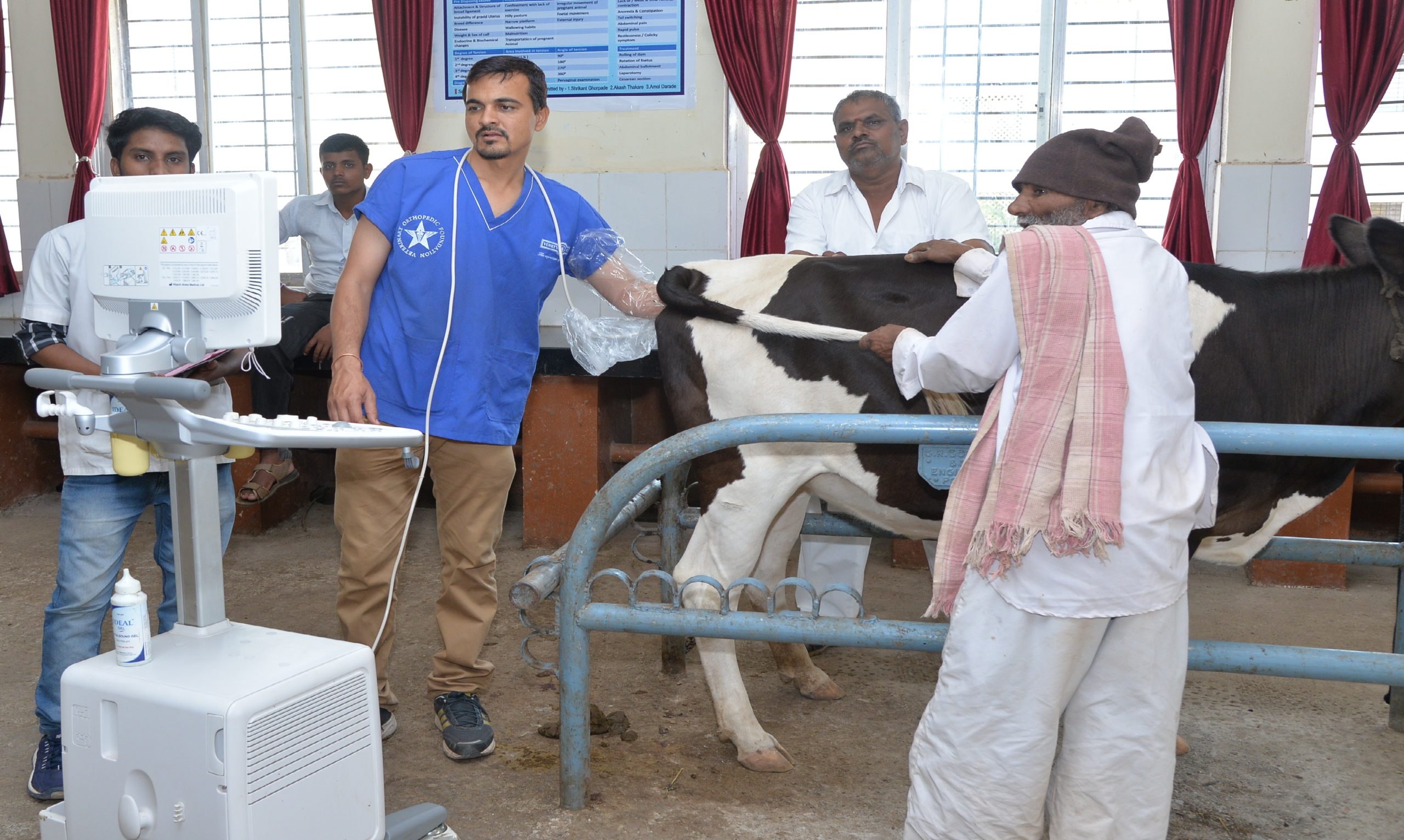
30 Days Pregnancy Diagnosis Concept in Dairy Animals
Dr. Ajit Mali,
Assistant Professor, ARGO, TVCC
KNP College of Veterinary Science, Shirwal, Maharashtra
Corresponding author email:- ajitvet7@gmail.com
Mobile No. : – 9850070481
Introduction:-
The milk productivity of dairy animals in India is comparatively quiet low. Two major important aspects that directly related to productivity of dairy animals are health and reproductive efficiency. Health of the animals can be maintained properly by using scientific animal husbandry practices such as vaccination, deworming, balanced nutrition and regular health monitoring protocols. Reproductive efficiency is tough job for Indian dairy farmers and veterinarians because it’s multifactorial origin and lack of awareness among farmers. Age of puberty, post-partum resumption of ovarian activity and ideal inter-calving period are important aspects to be focused by farmers and veterinarians to enhance reproductive efficiency. Our major task is to enhance size of breedable population and to reduce inter-calving period in prevailing livestock population. Infertility and repeat breeding are two major hurdles in improving reproductive efficiency in cattle and buffaloes. Early diagnosis of pregnancy as well as non-pregnancy plays crucial role in reducing infertility problems in farm animals. Dissemination of advanced diagnostic techniques to field veterinarians for management of reproductive health is highly essential. Real time Ultrasonography plays major role in reproductive health aspects in farm animals. Major advantages of ultrasonography techniques are simple, 100 percent accuracy, non-invasive, safe to animals as well as operator and require very less recurring cost for regular use. To enhance the reproductive efficiency in farm animals 30 days pregnancy diagnosis concepts by using ultrasonography should be adopted in dairy animals under field conditions.
Why Ultrasonography is need of the day?
Despite its numerous and relevant applications, only 20% of bovine clinicians across the world and very few in India use the ultrasound machine. Furthermore, many of those performing ultrasonography only use the scanner for detection of early pregnancy and research purpose in veterinary universities across the country. The constant application of ultrasonography can lead to a more accurate use of drugs as per the specific diagnosis. It can also reduce, or even cancel, the normal range of error of manual palpation: when an excellent clinician manually examines the ovaries, the range of error is estimated to be of 30-35%, while it can rise up to 70% when physiological and pathological conditions are investigated. This can lead to a remarkably longer calving-conception interval in Bovines, as well as a more intensive use of drugs (hormones). This means a reduction in the number of open days (management costs of each non-pregnant cow or buffalo around Rs.300/day after 100 days post partum), the amount of drugs used and the cost of labor, leading to a total neat saving of Rs.8000 to 9000/- per milking cow or buffalo per month only. Hence, it seems clear that ultrasonography is an ideal tool for reproductive health management in Bovines. So there is need to incorporate Ultrasonography machine in each veterinary clinic in India.
Applications of Ultrasonography in Dairy Animal Practice:-
Ultrasonography was introduced to the field of veterinary reproduction to be primarily used in mares for early pregnancy diagnosis, detection of twins, and photographic documentation of pregnancy. Now, the technology has been extended to most of the common animal species. Some of the specific clinical uses are
- Determining the seasonal status of the ovaries.
- Determining whether the female has reached puberty.
- Monitoring follicles for diagnosis or assessing treatments.
- Monitoring the corpus luteum.
- Estimating the stage of estrous cycle.
- Differentiating luteal persistence from anovulatory conditions.
- Evaluating the time of breeding.
- Collecting follicular oocytes by trans-vaginal aspiration.
- Evaluating an animal’s potential to serve as embryo transfer recipient.
- Detecting and studying the early embryo and ageing of embryo.
- Assessing fetal viability.
- Evaluating post partum involution.
- Diagnosing ovarian pathologic conditions such as luteal and follicular cysts, peri-ovarian cysts, ovarian tumors, and hemorrhagic follicles.
- Diagnosing pathology of the tubular organs such as hydrosalphinx, pyometra, uterine cysts, collection of intra-luminal uterine fluid and fetal debris.
How to Perform Ultrasonography in Large animals:-
- Trans-rectal technique (Linear probe having frequency in between 5 to 8 MHz):
- Restraining the animal for trans-rectal ultrasound is similar to preparing for rectal palpation.
- Anal area is lubricated with the coupling gel prior to insertion of the hand.
- The transducer is dipped into the gel. Since the rectal wall is usually moist the use of a coupling medium may not be so important for trans-rectal examinations. However, better contact may be obtained when a coupling medium is used, especially when the fecal matter is dry and hard.
- While introducing the lubricated hand and transducer into the rectum, the same precautions used during rectal palpation must be taken to minimize rectal tears.
- Since, fecal material can cause distortions on the ultrasound image; it should be removed prior to examination.
-
- Special care must be taken to ensure that the transducer is advanced within the lumen of the rectum and not into a blind pocket which can be done by placing the fingers beyond the tip of the transducer during major forward movements.
Schematic diagram of trans-rectal scan in large animals
- Trans-Abdominal technique (Convex sector probe having frequency in between 2.5 to 3.5 MHz):
- Trans-abdominal ultrasonography is usually performed with the animal in a standing position during mid or late gestation to know the status of fetus such as fetal movements, fetal heart beats, umbilicus blood flow and placentomes integrity.
- The convex transducer having frequency between 2.5 to 3 MHz should be placed in the inguinal region just cranial to udder of the cows or buffaloes.
- To eliminate the presence of air and promote contact between the transducer and skin, the hair of the inguinal area may be clipped in some breeds and the probe may be abundantly covered with contact gel.
- By trans-abdominal approach, mostly used during mid or late gestation because fetus moved in abdominal area during this period and it’s best approach rather trans-rectal scanning to monitor the fetus and related parts of pregnancy.
Evaluation of status of reproductive tract in dairy animals:-
- Uterus: –
- During trans-rectal ultrasonography, the first structures to be evaluated are the body of the uterus and the uterine horns.
- The non-gravid, fully involuted, uterus is a muscular structure and generates an echogenic image whose echogenicity depends on the uterine tone and luminal contents. Therefore, echogenicity differs during the luteal and follicular stages of the estrous cycle.
- The endometrial wall thickness can be measured and it is useful in differentiate whether animal is having subclinical endometritis or not. Generally endometrial wall thickness is up to 8 mm in normal cyclic animals and if it increases more than 8 mm and texture of endometrium become hyper-echoic then it can be easily diagnosed as subclinical endometritis.
- Ovary: –
- The ovaries appear elliptic, round or almond shape with a hyper-echoic outline. The diameter of ovaries is around 15 × 35 mm depending on the reproductive stage of animals.
- Identification of ovarian structures depends on the expertise and experience of technicians. Generally follicles are identified as anechoic round structure as it contains fluid whereas corpus luteum gives slightly hypo-echoic texture due to temporary tissue part of the ovary.
- In anoestrous females, the ovary is small and contains follicles between one and five millimeters. Owing to the fluid in the antrum, follicles are identified as black structures with a smooth spherical outline.
- The diameter of a follicle is measured by freezing the image and placing the electronic calipers of the ultrasound machine in the borderline between the follicular wall and the ovarian stroma.
- The ovulatory size of graafian follicle (GF) ranging from 11 to 18 mm in cows and buffaloes. The size of GF varies depending on parity and breed of animals.
- Pathological conditions in the ovaries such as, Cystic Ovarian Follicle (COF) both of luteal cyst or follicular cyst origin can be easily diagnosed and confirmed by ultrasonography.
- COF of Luteal cyst origin is mostly single with diameter of more than 20 mm with cyst wall thickness is more than 3 mm whereas in follicular cyst either single or multiple cyst with diameter of more than 20 mm of each cyst and cyst wall thickness is less than 3 mm.
Early Pregnancy Diagnosis in Dairy Animals:-
The use of trans-rectal ultrasonography to assess pregnancy status early during gestation is among the most practical applications of ultrasound for dairy animal reproduction. Early identification of non-pregnant dairy animals post-breeding improves reproductive efficiency and pregnancy rate in cows and buffaloes by decreasing the interval between AI services and increasing AI service rate. Under most on-farm conditions, pregnancy diagnosis can be rapidly and accurately diagnosed using ultrasound as early as 26 day post AI. Although pregnancy status can be established early, care must be taken to ensure the accuracy of a diagnosis. Ultrasound is a rapid method for pregnancy diagnosis, and experienced palpators adapt to ultrasound technology quickly by detecting embryonic vesicle, embryo proper with heart-beats, amniotic membrane and corpus luteum verum on day 30 post-breeding. Also embryonic or fetal ageing can be done my taking measurement of crown rump length and skull diameter of embryo from day 30. Concept behind 30 days pregnancy diagnosis in dairy animals is that estrus interval of 21 days, if animals not show post AI estrus after 21 days can be scanned by ultrasonography on day 30 of gestation. If animal is pregnant on day 30 well and good but if animal is non-pregnant then on the basis of ovarian and uterine status, estrus induction can be carried out by either hormonal or non-hormonal regimens. So by using 30 days pregnancy diagnosis concept we could get earliest detection of pregnancy or non-pregnancy status with any pathological conditions of ovary or uterus, less and accurate drugs requires for estrus induction which reduces the cost of treatment as well as time interval.
Conclusion:
Thus Ultrasonography technique is very easy, safe, economical and practical way to confirm reproductive status either pregnant or non-pregnant from 25 days of gestation in dairy animals. Also it determines either single or twins pregnancy with gestational age in animals whom history of breeding or AI is unknown. The ultrasonography is used as a diagnostic as well as applied research tool. In the veterinary imaging world, it is hard to beat ultrasound as it is safe, wide range of applications and cost-effective. Ultrasonography becomes the preferred diagnostic imaging modality of the 21st century in the clinical field of Animal Reproduction. The scanner will never replace the vet, but can be a simple, clear tool that helps him to diagnose and reduce the range of the error. To enhance the reproductive efficiency in farm animals 30 days pregnancy diagnosis concept should be adopted in dairy animals under field conditions in India.
References:
- A. B. Mali, M. V. Ingawale, N. M. Markandeya and B. L. Kumawat. (2019). Ultrasonographic studies on early embryonic developments and ageing in Murrah buffaloes under farm conditions, International Journal of Livestock Research, Vol 9 (01) : 271-277.
- A. B. Mali, S. R. Chinchkar, S. U. Gulavane, S. A. Bakshi and H. S. Birade. (2019). Ultrasonographic Diagnosis of Early Pregnancy in Buffaloes, Haryana Vet. 58 (S.I), 40-42.
- A. B. Mali, S. R. Chinchkar, S. U. Gulavane, S. A. Bakshi and H. S. Birade. (2018). Comparative study of early pregnancy diagnosis in buffaloes using ultrasonography and serum progesterone profile, Intas Polivet, 19 (II): 351-353.
- A. B. Mali. (2015). Application of Ultrasonography in Field Conditions for Improving Reproduction in Bovines, Intas Polivet, 16 (I): 11-15.


