Canine Demodicosis: An overview
Sneh Lata Chauhan*, Divya Agnihotri, Tarun Kumar, Neelesh Sindhu and V.K.Jain
Department of Veterinary Clinical Complex, LUVAS, Hisar, Haryana
*Corresponding author:
Dr.Sneh lata Chauhan (Scientist)
Incharge, Skin Disease laboratory
Veterinary Clinical Complex, LUVAS, Hisar, Haryana
Introduction:
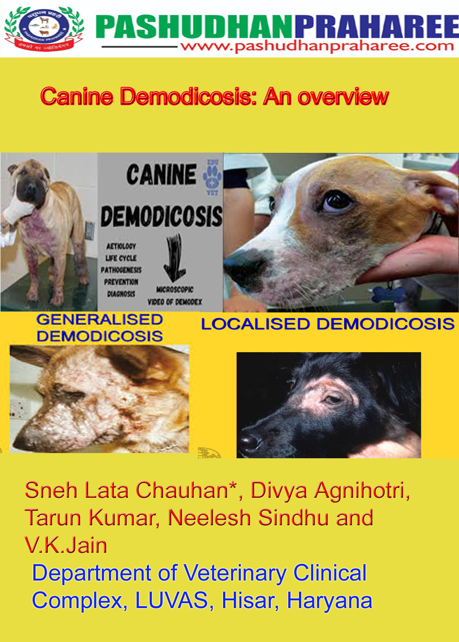
Canine demodicosis is one of the more common skin diseases encountered in veterinary practice. Demodex canis is a normal member of cutaneous ecology of the dog, but in some situations overpopulates resulting in skin disease. Two forms are recognized, a localized form and a generalized form. Localized demodicosis occurs most commonly in young dogs, less than a year of age. Typical lesions are erythematous and alopecic patches on the head and/or forelegs. Pruritus and fine scaling may be present. Spontaneous remission occurs in most patients. There is no uniformly accepted definition of localized vs. generalized demodicosis. The author considers involvement of an entire body region, more than five focal areas and/or paw involvement indicative of generalized demodicosis. The mite demodex canis is an ectoparasites and a normal inhabitant of canine hair follicles and sebaceous glands of the skin. Mites are transmitted by direct contact between the dam and puppies shortly after birth. Canine demodicosis occurs when an alternate immune response allows over proliferation of mites, leading to clinical signs..( Mueller; 2004)
Etiology:
The three recognized canine Demodex mites are: Demodex canis, Demodexinjai, and the unnamed short-bodied mite. Demodex canis was the first to be identified and named the two additional Demodex mites may be mutations of Demodex canis, or separate species. Hillier and Desch (1997) described Demodex injai, a long bodies demodecid, where the male mites were more than twice the length of the males of D.canis (Desch and Hillier 2003). In another report an unusual mite was reported by Scarff (1988). Stubby form of the Demodex was described as being about one half of the length of the female of D.canis (Chesney 1999). Currently, the reports about D. cornei infestation in dogs are very few in India, although the first report was published in 1998 (Scott et al. 2001). This paper reports the occurrence of mixed Demodex infestation of D. cornei and D. canis in dogs and its management.
Types of Demodicosis:
Two types of demodectic mange commonly found in canines.
- Localized : – cases occur when these mites proliferate in one or two smalls, confined areas. This results in isolated scaly bald patches – usually on the dogs face creating a polka- dot appearance. Localized demodicosis is considered a common ailment of puppyhood, and approximately 90% of cases resolve with no treatment of any kind.
- Generalized: Demodectic mange, in contrast, affects larger areas of skin of dogs and entire body. Secondary bacterial infections make this a very itchy and often smelly skin disease. This form of mange could also be a sign of a compromised immune system, hereditary problem, endocrine problem or other underlying health issue.
The immune system plays a role in the development of juvenile and adult –onset demodicosis.
ADULT ONSETS: – generalized adult – onset demodicosis, which clinically can appear as squamous or pustular, has been produced using immunosuppressive in adult dogs in which genetic factors are unlikely. A natural decline in on-specific immunity in older dogs can incite the emergence of demodex.
JUVENILE ONSET: -when the case is presented below one year of age. It can be localized and generalized.
Clinical Features:
- Many patients present with circular areas of alopecia. Occasionally, patients present with a diffuse area of thin hair coat.
- Pruritus is usually absenting unless a concurrent allergy or secondary skin infection develops.
- If untreated, these patients may also develop hyperpigmentation and lichenification along with increase body odor due to excess sebum production from sebaceous glands associated with hair follicles.
- Draining tract may also form due to rupturing hair follicles.
- Dogs can have systemic illness with generalized lymphadenopathy, lethargy, and fever when deep pyoderma, furunculosis or cellulitis is seen.
Diagnosis:
- Skin scraping: to find mites, it is necessary to obtain multiple, deep skin scrapings from affected areas. Scraping should be deep enough to produce capillary bleeding while squeezing the area being scraped
- Microscopy: fusiform eggs, six – legged larvae, eight –legged larvae, eight legged nymphs or eight legged adult mites
- Trichography: Trichography may be used to search for adult mites attached to the hair shaft. This procedure involves plucking some hairs in the direction of hair growth and placing them in mineral oil on a glass slide for microscopic examination. Because trichography is not as reliable as skin scraping for diagnosing demodicosis. It should complement and not replace skin scraping. Trichography may be helpful for yielding mites when collecting samples from areas of the skin that are difficult to squeeze or scrape, such as interdigital and periocular areas. Negative results from trichography do not rule out a diagnosis of demodicosis. (Tater & Patterson 2008)
- Polymerase Chain Reaction
Treatment:
- Application of benzyl peroxide or hydrogen peroxide containing preparation like shampoo for at least 15 min. Bathing with a benzoyl peroxide shampoo before dipping may be beneficial for its keratolytic effect and follicular flushing activity. Only after dipping in such shampoo amitraz is applied.
- Amitraz: – it improves the prognosis of the disease. The person applying the amitraj dip should wear gloves and protective clothing, and the patient should be treated in a well-ventilated area.
- Recent advances in the systemic treatment of generalized canine demodicosis indicate that daily oral administration of ivermectin, milemycine oxime, and moxidectin are very effective.
- Amitraz rinses at 0.025 -0.06 % every 7-14 days, and oral daily ivermectin at 300 ug kg -1, milbemycin at 2mg/ kg
Monitoring Response to Treatment:
After treatment begins, patients should be checked every 4 to 6 weeks until two consecutive negative skin-scraping results are obtained. By the first recheck examination, very few, if any, Demodex mites in immature stages should be seen. Ideally, the percentage of dead adult mites should be higher than the percentage of live adult mites. This indicates that the owner is being compliant and the therapy is working. Treatment should be continued for 4 weeks after the second negative skin-scraping result. Rechecks and skin scrapings should be performed every 3 to 4 months for the next year. Relapses can occur within this time frame, so owners need to observe their dogs carefully for evidence of disease. One year after a second negative skin-scraping result and no recurrence of disease, the patient can finally be declared cured.
References:
- Mueller, Ralf S. “Treatment protocols for demodicosis: an evidence‐based review.” Veterinary dermatology2 (2004): 75-89.
- Scott DW, Miller WM, Griffin CE. Parasitic skin diseases. In: Di Berardino C, editor. Muller and Kirk’s small animal dermatology. 6. Philadelphia: W.B. Saunders Company; 2001. pp. 423–516.
- Desch CE, Hillier A. Demodex injai: a new species of hair follicle mite (Acari: Demodecidae) from the domestic dog (Canidae) J Med Entomol. 2003;40(2):146–149. doi: 10.1603/0022-2585-40.2.146.
- Hillier A, Desch CE. A new species of Demodexmite in the dog: a case report. Tennessee: Annual Members Meeting of the American Academy of Veterinary Dermatology and the American College of Veterinary Dermatology Nashville; 1997. pp. 118–119.
- Tater KC, Patterson AC. Canine and feline demodicosis. Vet Med 2008:444-461.


