SURGICAL MANAGEMENT OF EPITHELIOGENESIS IMPERFECTA WITH CONGENITAL EVENTRATION IN A NEONATAL CALF
Dr.S.Tina Roshini
Assistant Professor, Rajiv Gandhi Institute of Veterinary Education and Research
Puducherry-605009
ABSTRACT
A five hours old female crossbred jersey calf was presented to the Department of Veterinary Surgery and Radiology of Rajiv Gandhi Institute of Veterinary Education and Research with eventration of intestine through a ventral abdominal defect since birth. There was no development of skin in the ventral abdominal region around the defect. Clinical examination revealed subnormal temperature with difficulty in breathing. The calf was stabilised by removing the mucus from the nostrils and by administering warm saline with dexmetasone at the dose of 0.1mg/ kg body weight. The calf was placed in dorsal recumbency and the ventral abdomen was surgically prepared. The surgical area was anesthetised by local infiltration of 2% lignocaine and the abdominal opening was extended cranially and caudally. The inflamed peritoneal layer covering the intestines was excised exposing the intestinal contents. The contents were pushed back into the abdominal cavity and the opening was closed by overlapping suture pattern using polyamide size 0. The subcutaneous tissue was closed using polyglactin 910 and skin by horizontal mattress using silk. Post operatively cefotaxim was administered for 5 days at the dose of 10 mg/kg body weight. The animal made an uneventful recovery.
Key words: Epitheliogenesis imperfect, congenital abnormalities, eventration
Introduction
Congenital malformations or congenital deformities are defined as abnormalities of structure present at birth (Badaway, 2011). The cause of many congenital defects is unknown, but some are inherited. Inherited disorders in cattle are mostly caused by autosomal recessively inherited genes (Janmeda et al., 2014). Epitheliogenesis imperfecta is defined as a condition where some areas of the body are devoid of skin at the time of birth (Benoit- Biancamano et al., 2006). Congenital eventration is defined as protrusion of visceral organs with its serous sac from faulty closure of the abdominal wall in the prenatal development life (Veena et al., 2011).
Case history and Observation
A five hours old female crossbred jersey calf was presented to the Department of Veterinary Surgery and Radiology of Rajiv Gandhi Institute of Veterinary Education and Research with eventration of intestinal contents through a ventral abdominal defect since birth (Figure 1). The contents were covered by peritoneal sac which was congested and thickened. There was no development of skin in the ventral abdominal region around the defect (Figure 2). Clinical examination of the calf revealed subnormal temperature of 96.4 F and difficulty in breathing. The calf was stabilised by removing the mucus from the nostrils and administering warm saline with dexamethasone at the dose of 0.1mg/ kg body weight. Otherwise the animal showed normal suckling reflex, defecation and urination. Once the rectal temperature increased to 97.9 F, the area around the defect was surgically prepared.
Treatment and discussion
The calf was placed in dorsal recumbency and 2% lignocaine at the dose of 2mg/kg was infiltrated around the surgical site (Figure 3). The abdominal opening was extended cranially and caudally (Figure 4). The inflamed peritoneal layer covering the intestines was incised and resected exposing the intestinal contents. The contents were pushed into the abdominal cavity and the defect was closed by overlapping suture pattern using polyamide size 0 (Figure 5 & 6). The subcutaneous tissue was closed using polyglactin 910 size 0 and skin by horizontal mattress using silk (Figure 7). Post operatively the calf was administered Inj. Cefotaxim at the dose of 10mg/kg body weight for five days intravenously, Inj. Meloxicam at the dose of 0.2 mg/kg body weight for three day intramuscularly and with oral multi vitamins syrup. The sutures were removed on the tenth day and the animal made an uneventful recovery. According to Leipold and Dennis, 1987 an estimated 0.5–1.0% of calves born have spontaneous congenital defects. Congenital ventral abdominal defects are very common in calves and these defects in the development of somatopleura leads to various defects in the bodywall especially in the ventral median parts (Veena et al., 2011). Giggin et al. 2022 reported congenital eventration with epitheliogenesis imperfecta in a day-old kid (capra hircus) and similar surgical management. Senna et al. (2003) also reported epitheliogenesis imperfecta with visceral eventration through umbilicus in lamb and kid and its surgical correction. Niwas et al., 2020 reported a case of eventration in a calf with soiled intestinal contents and on surgical correction that lead to peritonitis. Hence soiled intestinal visera may have negative effect on the prognosis of the calf.
Conclusion
Congenital malformations are common in ruminants and can be avoided by preventing inbreeding. Prompt surgical management has to be advocated to improve the quality of life for the affected calves.
Reference
- Badawy AM (2011).Some congenital malformations in ruminants and equines with special reference to the surgical treatment of recto–vaginal and cysto–rectal fistulae. Benha Vet. Med. J., 1:14-27.
- Benoit-Biancamano, M., Drolet, R. and D’Allaire, S. (2006). Aplasia cutis congenita (epitheliogenesis imperfecta) in swine: observations from a large breeding herd. Vet. Diagn. Invest. 18: 573–579.
- Giggin T., Deny J., Anoop S., Sudheesh S. N., Soumya R. and John Martin K. D. (2022). Congenital eventration with epitheliogenesis imperfecta in a Day-Old Kid (Capra Hircus) and its surgical management. Indian Vet. Assoc., 20 (2): 81-84.
- Janmeda M, Pandya G.M, Dangar N.S and Kharadi V.B. (2014). Congenital defects in cattle. Indian Farmer., 1(1): 15-18.
- Leipold HW, Dennis SM. (1987). Congenital defects of the bovine central nervous system. Vet Clin North Am Food Anim Pract. 3:159–177.
- Senna, N.A., Abu-seida, A.M., Gadallah, S.M., El-husseiny, I.N. and Rakha, G.M. (2003). Congenital anomalies in native breeds of sheep and goats: A report on 120 cases of 24 varieties. Vet. Med. J. 3: 363-380.
- Veena P, Sankar P, Kokila S, Kumar RVS, Lakshmi DN (2011). Congenital umbilical defect with visceral eventration in a buffalo calf – A case report. Buffalo Bulletin, 30:165-167.
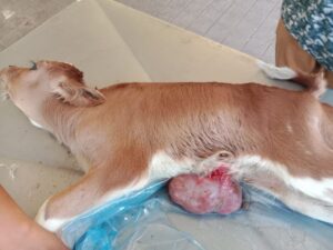
Figure 1: Calf with eventration of intestine with epitheliogenisis imperfecta |
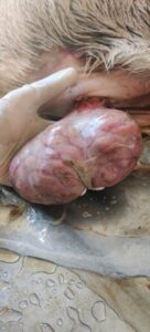
Figure 2: peritoneal sac covering the intestine |
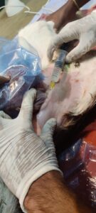
Figure 3: Infiltration of local anaesthesia |
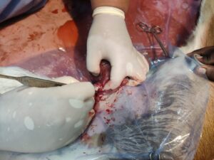
Figure 4: Extention of abdominal opening |
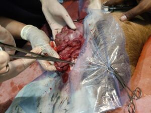
Figure 5: Reposition of intestine into the abdomial cavity |
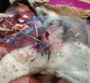
Figure 6: Closing of abdominal defect |
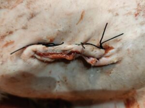
Figure 7: closure of skin |
|

