GASTROESOPHAGEAL REFLUX DISEASE IN DOGS
Varun Kumar Sarkar*, Harshit Saxena, Pooja Solanki*, Rajashekhar Kamalla* and Vandana*
*Division of Medicine, ICAR- Indian Veterinary Research Institute, Izzatnagar, Bareilly (UP)
Abstract
Gastroesophageal reflux is a normal physiological process and gastroesophageal reflux disease develops when the body’s natural anti-reflux barrier fails to adequately protect against frequent and high levels of gastric reflux. The classical clinical observations include repeated lip-smacking, drooling, an increase in empty swallowing motions, sudden unexplained discomfort, chronic intermittent vomiting, excessive grass eating, star gazing behavior etc. A telemetric pH measurement technique was used to diagnose gastroesophageal reflux disease along with Gastroduodenoscopy and histopathology to confirm the diagnosis. Nowadays Impedance planimetry and nuclear scintigraphy was used to diagnose the disease. Proton pump inhibitors like omeprazole or pantoprazole are still considered as the mainstay treatment for gastroesophageal reflux disease, however GABAB agonist baclofen and mavoglurant a selective allosteric modulator of mGluR5 are novel drugs used to treat gastroesophageal reflux disease. Whereas electrical stimulation is a new approach for treating gastroesophageal reflux disease patients who are dissatisfied with pharmacological therapy and apprehensive about the possible hazards of laparoscopic anti-reflux surgery.
Introduction
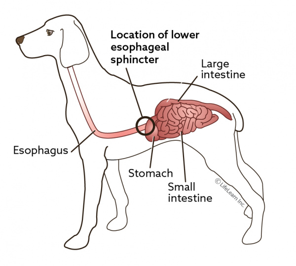
The esophagus is a muscular hollow tube with three functional regions: the upper esophageal sphincter (UES), the tubular section of the esophagus, and the lower esophageal sphincter (LES). As an esophageal barrier, the UES guards against stomach reflux and aspiration. According to research utilizing UES manometry, the adult UES is 2.5 to 4.5 cm long. In the healthy individual, a high-pressure zone between the stomach and esophagus is formed more distantly by the LES and the crural diaphragm. This zone serves to stop the reflux of gastric contents into the esophagus (Pitt et al., 2017). The condition known as gastroesophageal reflux disease (GERD) is brought on when the body’s normal anti-reflux barrier is unable to defend against frequent and excessive levels of gastroesophageal reflux (GER). According to Paran et al. (2023), dogs have a 50% prevalence of GERD. A number of times a day, GER occurs naturally as a physiological mechanism. GERD is thought to be a complex process, and it can cause discomfort, inflammation, and regurgitation. It can also cause typical reflux-related oesophageal consequences, such as ulcers, strictures, and epithelial metaplasia. Additionally, respiratory issues including laryngitis, bronchial irritation, and aspiration may occur. Clinical manifestations may also disappear after therapy with gastric acid inhibitors (Muenster et al., 2017; Kook et al., 2014). The method for diagnosing GERD in dogs has recently improved and now includes the use of telemetry equipment designed for humans to detect gastro-oesophageal reflux (Kook et al., 2014). Proton pump inhibitors (PPIs) are still the cornerstone of pharmacological management for GERD, with antacids or histamine type-2 receptor antagonists serving as backup options. Although PPIs have been demonstrated to be safe and effective in treating reflux esophagitis (Rouzade-Dominguez et al., 2017).
Etiology
- Young Shar Peis are most frequently reported to have sliding hiatal hernias as congenital anomalies, but it can also develop as an acquired syndrome as a result of airway obstructive disease or neuromuscular conditions that affect the diaphragm. It frequently coexists with GER (Mayhew et al., 2017).
- Momentary LES relaxations without causing any anatomical disturbance.
- LES hypotension, which occurs without consideration of anatomical abnormalities.
- Esophagogastric junction anatomical deformity, including hiatus hernia.
- Several anaesthetics may reduce the tone of the LES and increase the risk of GER while under anaesthesia.
- Prolonged fasting and intra-abdominal operations both increase the risk of GER (Poirier-Guay et al., 2014).
Pathogenesis
GERD is caused by a chemical insult to the squamous epithelium’s surface of the esophagus lumine that travels through the epithelium and lamina propria into the submucosa, destroying surface cells by acid infliction and inducing a proliferative response in basal cells. The updated pathophysiology of GERD in dogs is that refluxed gastric juice induces oesophageal epithelial cells to proliferate and release chemokines such as interleukin-8 and interleukin-1beta rather than directly damaging the oesophageal mucosa. This attracts and activates immune cells, which destroy the adult squamous oesophageal epithelial cells. Thus, surface erosions are preceded by basal cell and papillary hyperplasia. This updated model of GERD development in dogs may help to explain epithelial hyperplasia in the absence of erosions. It appears that epithelial hyperplasia in dogs is less severe than in humans with GERD (Muenster et al., 2017). It’s possible that during a reflux episode, the canine oesophageal mucosa is less exposed to acid (Kook et al., 2014), and that Barrett’s esophagus and other signs of severe chronic acid exposure, such as dilated intercellular spaces, are less common in dogs (Muenster et al., 2017).
Clinical finding
Historical observations noted in the medical records include repeated lip-smacking, drooling, an increase in empty swallowing motions, sudden unexplained discomfort, presumed postprandial pain, chronic intermittent vomiting, excessive grass eating, refusal to eat despite interest, regurgitation, retching, chronic mild intermittent cough, halitosis, and excessive surface licking, aspiration pneumonia, coughing, snoring, sneezing, weakness, lethargy, odynophagia, syncope, dysphagia, weight loss, stertor, gagging after drinking (Mayhew et al., 2017) and star gazing behavior (Poirier-Guay et al., 2014). On physical examination, laryngitis and tonsillitis were also discovered. Additional symptoms were concurrent intermittent small intestinal diarrhea (Kook et al., 2014).
Diagnosis
A diagnosis of GERD should be based on more reliable criteria such as-
Telemetric pH measurement (Bravo pH Monitoring System)
Dogs were sedated after a 12-hour fast in order to insert a pH capsule with endoscopic assistance. As a result, the esophageal capsule was situated around 4-5 cm above the Z-line. Using vacuum suction and a lock and pin mechanism, the pH capsule was able to adhere to the mucosa in accordance with the manufacturer’s instructions. A 6-second sampling interval was used for telemetric pH measurement after the capsule was inserted (Kook et al., 2014). The computer program determined the total number of refluxes, the number of refluxes lasting longer than five minutes, the length of the longest reflux (minutes), and the percentage of time when the pH was below four (fraction time pH four %).
Hispathology
Oesophageal samples are histologically analyzed. Hyperplastic thickening of the basal cell layer and aberrant extension of the stromal papillae of the lamina propria, two histological abnormalities associated with hyper-regeneratory oesophagopathy (HRO) that are regarded pathognomonic for GERD. But in order to confirm a diagnosis, these modifications must be prominent in at least one tissue sample (Muenster et al., 2017).
Gastroduodenoscopy
Multiple regions of superficial erosions and mucosal erythema in the mid and distal oesophagus were seen during a gastroduodenoscopy. At the gastroesophageal junction, the mucosa of the distal oesophagus had a notably erythematous and inflammatory appearance (Poirier-Guay et al., 2014).
Impedance planimetry
A method for obtaining in-flight recordings of the geometric details of gastric lumen architecture is impedance planimetry investigation. The commercially available EndoFLIP device comprises a generator with a syringe pump that allows regulated injection of a saline solution with known conductivity and a balloon-tipped catheter linked to it. At each balloon fill volume (30 and 40 mL), measurements of the minimum diameter, cross-sectional area, intra-bag pressure, and LES length were taken (Mayhew et al., 2017). Both in preclinical and clinical situations, this device has been utilized to evaluate the morphology and distensibility of the LES. It has been determined that people with GERD, have more LES distensibility than healthy participants (Pitt et al., 2017).
Nuclear scintigraphy
Another diagnostic method that can confirm GER is nuclear scintigraphy. However, the scan solely assesses postprandial reflux and fails to provide data on pH (Poirier-Guay et al., 2014).
Treatment
Proton pump inhibitors (PPIs), such as omeprazole or pantoprazole (median dose 1·5 mg/kg/day, range 1·0 -2·0 mg/ kg/day), are still the mainstay of pharmacological treatment for GERD, with antacids or histamine type-2 receptor antagonists like cimetidine, ranitidine, and famotidine being used as a backup (Rouzade-Dominguez et al., 2017). Thus, a unique pharmacological therapy that lowers the frequency of transient lower esophageal sphincter relaxations (TLESRs) and reflux episodes brought on by gastric distention is an alternate therapeutic strategy. TLESRs and the frequency of reflux episodes are reported to be effectively decreased by the GABAB agonist baclofen. Mavoglurant, at doses of 50 mg and 400 mg Single oral dosages are a structurally unique, subtype-selective allosteric modulator of mGluR5 that don’t appear to be harmful to the liver (Rouzade-Dominguez et al., 2017)
Surgery interventions
Through a celiotomy, all dogs received hiatal plication, esophagopexy, and left-sided incisional gastropexy (Mayhew et al., 2017).
Electrical stimulation
A substantial proportion of GERD patients who are dissatisfied with pharmacological therapy and apprehensive about the possible hazards of laparoscopic anti-reflux surgery may benefit from electrical stimulation of the LES, a relatively new approach for treating GERD (Rinsma et al., 2014).
Conclusion
Gastroesophageal reflux disease is a common clinical issue that is associated with serious health consequences and a possible decline in quality of life. The prevention of Gastroesophageal reflux disease consequences depends on early diagnosis of symptoms. Changes in behaviour and technological advancements in acid suppression are still crucial to its therapy.
Reference
Kook, P.H., Kempf, J., Ruetten, M. and Reusch, C.E., 2014. Wireless ambulatory esophageal pH monitoring in dogs with clinical signs interpreted as gastroesophageal reflux. Journal of veterinary internal medicine, 28(6), pp.1716-1723.
Mayhew, P.D., Marks, S.L., Pollard, R., Culp, W.T. and Kass, P.H., 2017. Prospective evaluation of surgical management of sliding hiatal hernia and gastroesophageal reflux in dogs. Veterinary Surgery, 46(8), pp.1098-1109.
Muenster, M., Hoerauf, A. and Vieth, M., 2017. Gastro‐oesophageal reflux disease in 20 dogs (2012 to 2014). Journal of Small Animal Practice, 58(5), pp.276-283.
Paran, E., Major, A.C., Warren‐Smith, C., Hezzell, M.J. and MacFarlane, P., 2023. Prevalence of gastroesophageal reflux in dogs undergoing MRI for a thoracolumbar vertebral column pathology. Journal of Small Animal Practice.
Pitt, K.A., Mayhew, P.D., Barter, L., Pollard, R., Kass, P.H. and Marks, S.L., 2017. Consistency and effect of body position change on measurement of upper and lower esophageal sphincter geometry using impedance planimetry in a canine model. Dis Esophagus, 30(4), pp.1-7.
Poirier-Guay, M.P., Bélanger, M.C. and Frank, D., 2014. Star gazing in a dog: Atypical manifestation of upper gastrointestinal disease. The Canadian Veterinary Journal, 55(11), p.1079.
Rinsma, N.F., Bouvy, N.D., Masclee, A.A. and Conchillo, J.M., 2014. Electrical stimulation therapy for gastroesophageal reflux disease. Journal of neurogastroenterology and motility, 20(3), p.287.
Rouzade‐Dominguez, M.L., Pezous, N., David, O.J., Tutuian, R., Bruley des Varannes, S., Tack, J., Malfertheiner, P., Allescher, H.D., Ufer, M. and Rühl, A., 2017. The selective metabotropic glutamate receptor 5 antagonist mavoglurant (AFQ 056) reduces the incidence of reflux episodes in dogs and patients with moderate to severe gastroesophageal reflux disease. Neurogastroenterology & Motility, 29(8), p.e13058


