


ABOMASAL DISPLACEMENT IN CATTLE
Dr Anita Kumari and Dr Kavipriya Jaiswal
INTRODUCTION
The abomasum is suspended loosely by the greater omentum and lesser omentum, it can be moved from its normal position on the ventral part of the abdomen to the left or right side (LDA and RDA, or it can rotate while displaced to the right and lateral to the liver (abomasal volvulus). The abomasum can shift from its normal position to left displacement or to right displacement over a relatively short period (pendulous LDA). Abomasal volvulus can develop rapidly or slowly from an RDA. Left displaced abomasum (LDA) is displacement of the gas-filled, distended abomasum to the left side of the abomasum, trapping it between the rumen and the abdominal wall. Right displaced abomasum (RDA) is displacement of the gas-filled, distended abomasum from the ventral abdominal wall into the cranio-dorsal right abdominal cavity. Abomasal volvulus is rotation of organ on its mesenteric axis results in abomasal torsion or abomasal volvulus. LDA and RDA result in partial ileus; abomasal volvulus leads to complete ileus and abomasal wall ischemia.
ETIOLOGY
The etiology of displaced abomasum and abomasal volvulus is multifactorial. Decreased abomasal emptying by abomasal hypomotility and/or dysfunction of the intrinsic nervous system are thought to play an important role in development of displacement or volvulus. Important contributing factors include abomasal hypomotility associated with hypocalcemia and hypokalemia, concurrent diseases (mastitis, metritis) associated with endotoxemia and decreased rumen fill, Periparturient changes in the position of intra-abdominal organs, and Genetic predisposition, particularly in deep-bodied cows. Genetic predisposition is correlated with milk production, milk production increase the incidence of abomasal displacement. Hypomotility is also related to ingestion of high-concentrate, low-roughage diets, which reduce abomasal motility through a poorly defined mechanism that may involve hyperinsulinemia or increased concentrations of volatile fatty acids. Finally, subclinical and clinical ketosis increase the risk of abomasal displacement through an unknown mechanism that may be associated with decreased rumen fill. Approximately 80%–85% of displacements occur within 1 month after parturition; however, they can occur at any time, including during late pregnancy.
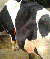
CLINICAL FINDINGS
- The typical history of abomasal displacement includes anorexia (most commonly a lack of appetite for grain with a decreased or normal appetite for roughage).
- Decreased milk production (usually notable but not as extensive as with traumatic reticuloperitonitis or other causes of peritonitis).
- In abomasal volvulus, anorexia is complete, milk production is more markedly and progressively reduced, and clinical deterioration is rapid.
- In abomasal displacement, temperature, heart rate, and respiratory rate are usually normal.
- The caudal part of the rib cage on the side of the displacement may appear “sprung.”
- Hydration appears subjectively normal with LDA except in some chronic cases; in contrast, in abomasal volvulus, dehydration occurs very early.
- Rumen motility may be normal but often is reduced in frequency and strength of contraction.
- Feces are usually reduced in quantity and more fluid than normal; however, they may be shed with normal consistency
DIAGNOSIS
- On the basis of History, clinical signs, auscultation and percussion.
- Clinical pathology
- Liptek test – An 18G needle is inserted aseptically just below the area of resonant ping in the left and or in right abdominal wall and fluid is aspirated. If the pH of fluid is between 1-4, an abomasal source should be suspected.
- The most important diagnostic physical finding is a ping on simultaneous auscultation and percussion of the abdomen, which should be performed in the area marked by a line from the tuber coxae to the point of the elbow, and from the elbow toward the stifle on both sides of the patient.
- The ping is characteristic of LDA is most commonly located in an area between ribs 9 and 13 in the middle to upper third of the left abdomen; however, the ping can be more ventral or more caudal, or both.
- The ping associated with RDA also is most commonly located in the area between ribs 10 and 13 on the right abdomen.
- A ping cranial to rib 10 usually indicates the presence of abomasal volvulus because the liver is displaced medially by the distended viscus.
TREATMENT
Conservative medicinal treatment
- To correct dehydration
- Correction of metabolic disorders
- Restoration of gastrointestinal motility
Surgical treatment
- Left flank omentopexy (Utrecht method)
- Left flank abomasopexy
- Right flank omentopexy
- Right pyloricantropexy (”pyloropexy”)
- Right paramedian abomasopexy
REFERENCES
Geishauser, T. (1995) Abomasal displacement in the bovine – a review on character, occurrence, aetiology and pathogenesis. Journal of Veterinary Medicine, Series A 42, 229-251
Gordon, P. (2009) Roll and toggle correction of left displaced abomasum (LDA) – practical tips for success. Cattle Practice 17, 128-130