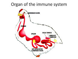
CONCEPT NOTES ON THE LYMPHATIC SYSTEM OF POULTRY
Post no 1370 Dt 24th August 2019
Compiled & shared by- DR RAJESH KUMAR SINGH, JAMSHEDPUR,9431309542,rajeshsinghvet@gmail.com.
Introduction:
Lymph is a colorless fluid which passes out of the blood into a network of fine tubes called the lymphatic system.It passes through the lymph nodes, where germs are filtered out and killed, before it is returned to the veins.The lymph nodes and spleen also produce special blood cells which protect the body against disease.Sometimes when an animal is infected the lymph nodes become swollen and can be felt beneath the skin.
Understanding the physiology and immunology of lymphoid system is handicapped without knowledge of its basic structure. The body of birds is richly served with lymph vessels. Lymph is derived from the body fluid found within the interstitial space (the space between the important parts of the body). It includes lymph, lymph vessels and lymph nodes. Generally. Lymph flows away from the tissue to lymph nodes and eventually to either the right lympatic duct or the largest lymph vessels in the body, the thoracic duct. The lymphatic system is a system which collects and returns the interstitial fluid. As with the blood network, the lymph vessels form system all through the body, contrast to the blood, in which the lymph system will be in one direction draining lymph from the tissue and returning it to the blood. This system is a network of capillaries and tubes called “Lymphatics”. These lymphatics use up lymph from all over the body. The lymphatic system protects the body against disease by producing lymphocytes. It is also absorbs lipids (fats) from the intestine and transports them to the blood. Lymphatics are found in every part of the body apart from the central nervous system. The major components of the lymphatic system are bone marrow, lymph nodes, spleen, the bursa of fabricus and the thymus gland. The lymphatic system of birds is characterized by a number of morphological features which place it phylogenetically in an intermediate position between the analogous system of amphibian & reptiles and that of mammals. There are no lymph nodes in fowls. Lymph plexuses (an interwining of the very small lymph vessels) are found instead of mammalian lymph nodes.In the duck and the goose, we recognize two pairs of lymphnodes i.e. the cervico-thoracic lymph nodes and the lumbar lymph nodes. The lymphnodicervicothoracales are inserted into the vas lymphaceumjugulare of each side. They are situated close against the jugular vein at either the caudal end of the neck or in the cranial part of the body cavity. They are 10-15mm long and 3-5mm thick and have an elongated, spindle shaped structure. The lymphnodes of the birds develop by adaptation of the wall of the relevant lymph vessel, which in this case is the vas lymphaceumjugulare. The Inn.Lumbales are also paired and of similar structure. They are situated immediately under the vertebral column and are incorporated in the ductusthoracicilumbales which accompanies the a. sacralis media. In goose, they measure some 2.5cm in length and 0.5cm in thickness. They extend between the point of origin of the external iliac artery and the ischiatic artery. The lymphatic system has the function of draining the body systems of fluid that is left behind by the blood vessels, although the lymph fluid does ultimately return to the main circulatory system, when the lymph vessels enter the venacava near the heart.In general, lymphatic system is poorly developed when compared with mammals. Unlike the cardiovascular system, the lymphatic system is not closed and has no central pump. The lymph movement occurs despite low pressure due to peristalsis (propulsion of the lymph due to alternate contraction and relaxation of smooth muscle), valves and compression during contraction of adjacent skeletal muscle and arterial pulsation.Anatomically lymphatic vessels or lymph vessels are thin walled, valve structures that carry lymph. Lymph vessels are lined by endothelial cells and have a thin layer of smooth muscle and adventitia that bind the lymph vessels to the surrounding tissue. Lymph vessels are devoted to propulsion of the lymph from the lymph capillaries which is mainly concerned with absorption of intestinal fluid from the tissues. Lymph capillaries are slightly larger than their counterpart capillaries of the vascular system. Lymph vessels that carry lymph to a lymph node are called the afferent lymph vessels and one that carries it from a lymph node is called the efferent lymph vessels from where the lymph may travel to another lymph nodes, may be returned to a vein or may travel to a larger lymph duct.
Function of lymphatic system:
i) It serves as system for draining tissue fluid. ii) It carries proteins and even large particulate matter away from the tissue spaces. iii) It helps in absorption and transportation of fats and vitamin K from the small intestine to the blood. iv) It distributes the digested food to the tissues and collects the metabolic waste substances. v) It assists in the control of infection.
Lymph:
i) It is a tissue fluid which has entered lymph capillaries. ii) It is a colourless fluid. iii) It is practically blood minus the RBCs iv) They originate as blind sacs between tissue cells as capillaries and collects fluid not absorbed by venous system. v) These vessels pass through lymph nodes and finally join cranial vena cava by thoracic duct.
Lymphnodes:
i) They are spherical, oval or bean shaped. ii) They are grey rosy in colour. iii) They vary in size from very minute bodies to the size of lemon. iv) The lymphnodes are covered by a connective tissue capsule. v) The capsule sends septa or trabeculae inside the node. vi) The node is divided into cortex and medulla, which contains large number of lymphnodes. vii) They serve as filters for the lymph. viii) They act as one of the first body defense against infection.
Thymus:
The thymus is derived from the endodermal pharyngeal pouch epithelium, as are also the parathyroid glands and the ultimobranchial body. In birds, the 3rd, 4th and 5th pharyngeal pouches proliferate distally and initially form a solid column of cells. The avian thymus lies parallel to the vagus nerve and internal jugular veins. Thymus decreases in size as the bird mature.On each side of the neck, there are 7-8 separate lobes, extending from the third cervical vertebra to the upper thoracic segments. Each lobe is encapsulated with a fine fibrous connective tissue capsule and embedded in adipose tissue. From the capsule, septae invade the thymic parenchyma and incompletely divide the lobe into lobules. The button or bean shaped thymic lobes reach a maximum size of 0.6-1.2 cm in diameter by 3-4 months of age before physiological involution begins.Lymphocytes migrate into the cellular mass from the ingrowing blood capillaries and the surrounding mesenchyme and these cells are then transformed into reticulum cells.
Connective tissue septa divide the thymus into lobules with a cortex consisting of a dense collection of lymphocytes and a medulla made up mainly of reticulum layers. The thymus of birds is embedded into completely isolated lobes and in the fowl, there are 6-8. In duck and pigeon, it is 5- 6 of the oval and round, flattened lobes. In young fowls, they measure 8-15mm in length, 7-9mm in breadth and 2-5 mm in depth. The weight of thymus of a hen of about one year of age is 1.50-4.75gm. The organ about is said to attain its maximum weight in young, actively laying fowls but even in 5 years old hens, it still weight about 2 gm. However, the thymus does involute and ultimately it completely disappears in old birds. The thymus is a lymphoepithelial organ and therefore plays an important part in the defense mechanism against infection. It is also believed to exert an influence on the development of other lymphoreticular organs.
Bursa of Fabricus:
The bursa of fabricus develops initially as a solid protuberance on the dorsal wall of the urodeum of cloaca. Only when the epithelium lined lumen develops and forms into a pouch or bursa, it communicates with the proctodeum. In a fowl of about 8 weeks of age, it is round or pear shaped and about 1.5-2cm in length and 0.8cm in width. In chicken, bursa of fabricus has the shape and size of a chest nut and is located between the cloaca and the sacrum. A slot like bursal duct provides a continuous and free communication between the proctodeum and the bursal lumen. The bursa reaches its maximum size at 8-10 weeks of age, then like the thymus it undergoes involution. By 6- 7 month, most bursaare heavily involuted.With the onset of sexual maturity, it regresses and finally disappears. Its lumen is lined by simple columnar epithelium. Numerous tubules arise from it and between them are lymphoid follicles whose number increases during development. Removal of this lymphoreticular organ leads to a reduction of the defense mechanism against infectious diseases. The bursa of fabricius is sometimes referred to as the “cloacal thymus”, for there are certain structural and functional resemblances between these two organs, both contain numerous lymphoid cells and are important in immunological mechanisms.
Spleen:
The splenic primordium first appears as a mass of mesenchymal cells in the 48 hrs embryo. In contrast to that of mammals, the avian spleen is not considered a reservoir of erythrocytes for rapid release into the circulation. Spleen has an important role in embryonic lymphopoiesis.At the time of hatching, the spleen becomes a secondary lymphoid organ which provides an indispensable micro-environment for interaction between lymphoid and non lymphoid cells. The contribution of the avian spleen to the immune system, as a whole may be more important than in mammals because of the poorly developed avian lymphatic vessels and nodes.The chicken spleen is a round or oval structure lying dorsal to and on the left side of the proventriculus. The spleen is surrounded by a thin capsule of collagen and reticular fibre, poorly developed connective tissue from the capsule. In fowl, it is round or egg shaped. In the aquatic birds, it is more triangular with a flattened dorsal and a convex ventral surface, in the pigeon, it is oval. The spleen weight of the fowl and duck weight 1.5- 4.5 gm, the goose 4-8 gm and that of the pigeon is 0.2-0.4 gm. It lies against the dorsal surface of the right lobe of the liver in a space formed dorsally by the gonad, ventrally by the liver and laterally by the gizzard.The normal spleen is about 0.75 inch in diameter, located near the gizzard in the body cavity. Histologically, it is composed of red and white pulp. The function of spleen include phagocytosis of worn out erythrocytes in red pulp. Lymphocyte production in white pulp and antibody production in both the red and white pulp.Structurally and functionally, the spleen belongs to the blood forming organs and at the same time, it is also an important component of the reticulo-endothelial system which serves as a defense against noxious substances. In the embryo, it produces both red blood cells (RBC) and white blood cells (WBC) and it retains lymphatic ability throughout life. The spleen also removes erythrocytes which are no longer viable from circulation. Some components of the blood pigments are passed to the liver for further while iron is deposited in the spleen in the form of ferritin.
Bone marrow:
In adult birds, the medullary cavity (cavummedullare) of the long bones and the medullary spaces (cellulaemedullares) of the spongy bone are only partly filled by the blood forming bone marrow. The first blood cells, and also the endothelial cells lining of the blood vessels arise from mesenchymal elements called angioblasts which are situated in the blood islands of the area vasculosa of the germinal vesicle and wall of the yolk sac. Other sites of blood formation during development are the liver, spleen and thymus but the bone marrow finally becomes the blood formation in the higher vertebrates. However, lymphocyte production in birds also takes place in all types of lymphatic tissue, including the lymph nodes in duck and Ugeese.


