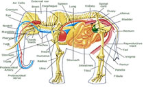
ANATOMICAL FEATURES OF ELEPHANTS
Understanding the anatomy of an elephant is the sine qua non for successful post-mortem of the animal. The following notes are meant to provide the field veterinarians with a basic understanding of the special anatomical peculiarities of elephants.
- Digestive system
- The proboscis, though a respiratory organ, acts as a prehensile organ to grasp the food and conveys it to the mouth for mastication.
- The lips are two musculo-membranous folds which surround the orifice of the mouth. The upper lip merges with the lower face of the proboscis. The lower lip is elongated with a pointed tip.
- The upper jaw has two incisors or tusks, one in each premaxilla. The tusks of the male grow to an enormous size in the adult Asian elephants. They are relatively much smaller in the female. There are male elephants which do not have tusks and are called Makhnas. Both cows and Makhnas possess tushes instead of tusks.
- The sub-lingual salivary gland and the sub-maxillary glands are present.
The tonsils are absent.
- The pharynx is a funnel shaped musculo-membranous sac common to both digestive and respiratory system. The soft palate divides the cavity into a dorsal large nasopharynx and a ventral small oesopharynx. The oesophagus communicates in front with the oral cavity and forms a deep diverticulum or receptacle (glosso-epiglottic space) behind the root of the tongue.
- Oesophagus, a thick walled musculo-membranous tube, is compressed dorsoventrally.
- The peritoneum, lining the abdominal cavity, is very thick and is reflected to cover the viscera. The greater omentum, which is one such reflection, is thin, lace like and devoid of fat. It is very extensive, attached to the greater curvature of the stomach and the origin of the duodenum.
- Stomach is a simple elongated musculo-membranous sac placed vertically behind the left part of the diaphragm and the liver.
- Small intestine is a mucous membrane forming longitudinal and transverse folds in honeycomb patterns.
- Diaphragmatic face of liver is strongly convex and the visceral face is deeply concave. The deep umbilical fissure divides the organ into a larger right and a smaller left lobe. The right lobe is extensive. The gall bladder is absent.
- Pancreas is dark brown, lobulated and situated in the mesoduodenum.
- Respiratory system
Proboscis is highly mobile, tactile and prehensile organ. It is made up of two nasal tubes separated by the septum nasi. The anterior margin of the tip of the proboscis has the “prehensile finger” which is highly tactile.
- The parietal pleura are very thick and adherent to the visceral pleura completely obliterating the pleural cavity. The parietal pleura are closely attached to the thoracic wall and are easily separable from the thoracic wall due to the abundance of the endothoracic facia.
- The left lung is smaller and extends from the 3rd rib to the 16th rib. Deep fissures mark the apical, cardiac and diaphragmatic lobes. The right lung is larger and extends from the 2nd rib to the 16th rib. Mediastinal lobe is attached to it.
- Cardiovascular system
- Heart is situated in the middle mediastinum extending frorm the 1st to the 5th rib with the apex resting on the 4th interchondral space. It is large, ovoid and longer on the left side.
- The apex of the heart in majority of the cases is bifid, incisura cordis separating the two apices of the ventricles
4. Urinary system
- The kidneys are multipyramidal, lobulated and covered by a dense layer of connective tissue and fat. The left kidney is placed ventral to the vertebral ends of the last four ribs. The dorsal face is convex while the ventral face is flat. The right kidney is larger than the left. It is situated under the vertebral ends of the last three ribs.
- Urinary bladder is small and pyriform lying at the pelvic inlet.
- Genital system
- Ovaries are oval and flat on the sides, situated in the abdomen, ventral to the iliac crest and posterior pole of the kidneys.
- Testes are situated on each side medial to the caudal pole of the kidneys. • There are two pectoral mammary glands. Each gland has a small conical teat. It is placed between the front legs with multiple openings.
- Haemopoietic system
- There are only a few lymph nodes in the mesentery.
- Hemal nodes are dark brown in colour, intercalated in the course of the blood vessels. These are more in number as compared to the lymph nodes.
- Spleen is in the shape of a dark and elongated organ, wide in the middle and pointed at both the ends. It is vertical in position and is related to the left abdominal wall The caudal border has a number of notches.
- Brain
- Elephant has a brain only four times the size of that of man. It has a large cerebral cortex.
- Skeletal system
Vertebrae — C 7, T 19-20, L 3-5
S 3-5, Cd 24-34
Ribs— 38 inl9 pairs (Sternal-6, Asternal-9, Floating ribs-4)
Compiled & Shared by- This paper is a compilation of groupwork provided by the
Team, LITD (Livestock Institute of Training & Development)
Image-Courtesy-Google
Reference-On Request.


