Alternative Technologies used for Anatomy Education
ABSTRACT
Use of animals for teaching is inevitable. Animals may be handled for anatomical demonstration, surgical procedure, experiments for various researches, pharmacology, physiology. To meet the purpose of learning and taking care of ethical issues in animals there is a need of an alternative way for teaching anatomy education which can stimulate students to conceptualize and develop easy understanding and learning of the topics. Earlier teaching of anatomy was thought to be “essential hands-on” that allowed students to engage all senses including the handling of instruments that allowed for the development of psychomotor skills, and the visualisation of structures. Visualisation in anatomy teaching is associated with a deep approach to learning. This approach is taken to next level by using various new techniques which have proven to be less traumatic to animals and have comparatively good shelf life.
The curriculum in teaching anatomy is under increasing pressure to transform from traditional to interdisciplinary integration, from cadaver-based to multimodal instruction with a system-based approach. Educational technologies are becoming critical and urged to be integrated into teaching medicine.
INTRODUCTION
The term anatomy is originated from the Greek word, ‘anatemnein’ where ana, means “separate, apart from” and temnein means “to cut up, cut open”.Whereas the meaning of the term anatomy is ‘Dissection’ in Latin.The traditional approach for teaching anatomy included use of dissection and large amount of cadavers but in due course of time the teaching and learning methods have evolved and changed. This includes use of various new technologies which have been launched in recent times to ease the understanding and learning of students. Such innovative technologies includes use of Veterinary anatomical term learning media, Surgical simulators, Visible Animal Project, Non-invasive methods such as Magnetic Resonance Imaging (MRI) and Computed Tomography (CT), 3D reconstruction of anatomical region of interest etc.These techniques use modern technology and virtual reconstructions for giving a realistic idea to the viewers. The dissection of cadaver had been gold standard of anatomy teaching and learning for a long time. Dissection requires an enormous number of cadavers, which puts a lot of strain on any source.But the computer-assisted learning, simulation-based training, using true-to plastic anatomical models, manikins, plastination are being used in recent times for both time and training costs advantages and to ensure healthy laboratory conditions. One of the reason of importance of anatomy as a subject for the veterinary education is that the knowledge of the combination of all subsystems of the body is required by the physicians so as to understand if there is a pathology in any part of the body or not. Various drawings of the internal organs of animals as well as external images on the walls are found on historical tracing of anatomical teaching process. Now-a-days the use of animals for teaching is minimized due to concern of suffering to the animals and more and more emphasis is given on the use of alternatives. Alternatives used for teaching and demonstration are durable, economical and does not involve killing or harming of living organisms. The alternatives become cost effective with due course of usage and time even if initially they are expensive. Few of the alternatives which are currently being used are discussed below.
As early as 2003, some methods for anatomy education were reported (Brenner et al.,5 2003). These education methods are in person- lectures, cadaver dissection, prosected specimens, models, radiological- living anatomy teaching and computer-based learning, for example, AR, VR, and 3D (Iwanaga et al.,6 2021).
Lectures
Anatomy lectures follows the learning objectives in which a lecturer presenting academic contents to a group of students. The traditional teaching lectures method requires the presence of the students in a specific location like lecture hall or classroom on precise time (Chang et al.,7 2019). Recently, student-centered policies such as flipped classroom, team-based learning and case based learning have shown to improve student collaboration, commitment and shift the focus of teaching from knowledge transmission to knowledge construction by the students (Bell et al.,8 2019). Engaging recent and alternate approaches in medical education has become crucial element today. Some anatomy classrooms use a wide range of technology in the form of e-textbooks, 3 D atlas, CD-ROMs, models and simulations(Singh et al.,3 2019).
3D atlas
3D atlas applications are designed as new learning materials in which applications are tablet-based software that permits students to touch and rotate the virtual bodies and recognize the spatial relationships. 3D atlas is not very helpful in memorizing the location of anatomical structures or gaining deep anatomical knowledge but it can simply help in the quick identification of the anatomical structures. The 3D atlas will show good link with 2D atlas if used properly in the gross anatomy (Park et al.,9 2019).
Plastic Models
Plastic models have been used to avoid problems associated with cadaveric dissection (McLachlan,10 2004). The plastic models are in common use in high schools and medical schools, but anatomical specimens are often lack essential specific details. Plastic models are suitable for some basic simple teaching purposes but not ideal for teaching detailed anatomy required in most of anatomy medical courses (McMenamin et al.,11 2014).
Simulation
Simulation is one of the educational methods implementing in the basic course of the anatomy (Torres et al.,12 2014). It is a technique, which substitutes or strengthens doctor and patient experiences in controlled settings and therefore evokes or repeats considerable aspects of the real world in a fully collaborative manner. Through simulated patients and peer examination, anatomy can be studied in the living body and this technique is very useful in studying bones, joints, muscles, peripheral nervous system and abdominal organs (Chang et al.,7 2019; McLachlan,10 2004).
Dissection
Cadavers have been always used in teaching anatomy and it is the gold standard simulator for the living anatomy (Darras et al.,13 2018). Cadaver dissection is the traditional method of teaching anatomy after theoretical classes and discussions on the atlas pictures. Some of the distinctive aspects of cadaveric dissection include the realistic environment of this teaching method that allows students to have and know clearly the organization of human body and experience the texture of the human tissues (Dissabandara et al.,14 2015). Cadaver dissection supports medical students in understanding the relationship of different anatomical structures, appreciating the anatomical variations and contributes significantly to a future professional work (Ghazanfar et al.,15 2018). Most students showed preference for having the choice to join dissection during their anatomy education (Whelan et al.,16 2018). In addition, surgical practices based on adequate anatomical knowledge of human anatomy which can be learned from cadaver dissection training contribute to an increase in surgical efficacy and self-confidence (Lim et al.,17 2018). Disappointingly, cadaver dissection is redundant from the anatomy curriculum in some medical schools due to shortage of cadavers, insufficiency of anatomists and short courses (Samarakoon et al.,18 2016). Dissection can necessitate recurrent access to dissecting rooms and laboratories by students which can be challenging to achieve mainly in institutions with limited resources. Furthermore, in some countries, dissection is prohibited due to religious (McMenamin et al.,11 2014). A review published of anatomical dissection as a teaching method in medical schools have been shown that a number of studies suggested dissection as a superior method of learning in comparable to non-dissection methods while some studies were of a conflicting opinion (Winkelmann,19 2007). Contradictory opinion from some students about cadaver dissection can be improved by preparing students effectively before the dissection sittings by using other methods of learning such as introductory lectures, prosection and model-based meetings prior to the dissection activities and by providing suitable guidance during the dissection sessions (Dissabandara et al.,14 2015).
Prosection
In the absence of cadaver dissection, prosected specimens (the use of pre-dissected cadaver specimens) have been widely used in the teaching anatomy to medical undergraduate students (Collins,20 2008). Interestingly, anatomical knowledge proceeding to prosection influenced the short term retention of the knowledge (Lackey-Cornelison et al.,21 2020).
Plastination
Plastination technique has been developed and is one of the best methods for preservation of organic tissue. It is widely used in anatomy to develop robust anatomical specimens of the whole body or body parts. In 1977, Dr. von Hagens in the Department of Anatomy at the University of Heidelberg planned plastination method for conserving anatomical specimens with reactive polymers (von Hagens,22 1979). In plastination technique, water and lipids in the biological tissues are replaced by polymers such as silicone, epoxy and polyester. After the hardening of these materials, odourless, dry, long-lasting and easily transported specimens were obtained (Sora et al.,23 2019). In sheet plastination, semi-transparent slices of tissue are obtained and students become easy to study the structural and topographical anatomy in detail (Sora et al.,24 2012). Plastination procedure offers fully preserved specimen without any foul smell or toxic fumes (Hayat et al.,25 2018). Specimens can be preserved and storage easily for longer times more than that of conventional method (Haque,26 2017). Plastination is not a replacement for traditional guided cadaveric dissection, but it does offer an additional learning implement to know and understand complex human anatomy (Riederer et al.,27 2014). Importantly, some students from recent studies have proved that the use of plastinated specimens is useful when learning anatomy (Latorre et al.,28 2016). Learning anatomy only in plastinated specimens is a compromise because of its restrictions in terms of tactile and emotional experience and skills that is delivered by wet cadavers (Fruhstorfer et al.,29 2011).
PLASTINATION involves the infiltration of specimens with synthetic materials and makes them of high durability. The water and fat of the body are replaced by certain polymers such as silicone resins or epoxy polymers.This method is used for long term preservation of the biological tissues with completely visible surface. The plastinates obtained after plastination retain properties of the original sample and avoids the decay of the specimen.These can be remodeled if misused.
FLASH BASED MODULES
These are designed to enhance the learning ability of the students and hones their learning skills.These help to refresh the knowledge on interdisciplinary anatomical features. The main advantage of use of modular teaching will be decreased usage of formalin, phenol, glycerol which are otherwise used for embalming and for storage of specimens. Other advantage is reduction in number of cadavers used for dissection. Being electronic in nature they provide chance of continuous revision of the topic thus leading to more memorization amongst the students as well as be useful to the practitioners or surgeons before going for any surgical intervention or operation so as to minimize the chances of error.
MODELS
Anatomical models are a great educational tool to study and explain the internal and external structure of the body. It refers to a smaller or larger physical copy of any live animal or organ system. Such models offer a three dimensional view to the observer and gives a realistic idea about the location and structure of certain organs. They can be made up from multiple materials ranging from rubber, plastic, POP, fiber, wooden and cemented.
All of the models have advantage that they are resistant to wear and tear and can be used for teaching and demonstration purpose for a good amount of time.
MANIKINS
It refers to the virtual patient which resembles the animal/ patient and can be used in the teaching and demonstration of CPR, first aid, tracheal intubation etc. it is also known as dummy/ mannequin/ life sized-dolls. SIMULATORS In simulators the visual components of the procedure are reproduced by computer graphic techniques. There is use of 3D CT or MRI scans of the patient data to enhance the realism inmedical simulators whereas the other web based simulations and procedural simulations can be viewed by the help of standard web browsers.
Body Painting
Body painting is used to assist the living anatomy, clinical skills classes and traditional anatomy courses. Several structures such as bones, muscles, vessels, nerves, and internal organs are painted on real living body and allowing for easy examination and palpation (Jariyapong et al.,30 2016). The costs are cheap and the entertaining activity can be concomitantly achieved with large classroom size. Using body painting exercise is helpful to promote active learning and increased knowledge retention whereby student’s pursuit for tools to accomplish understanding of the subject instead of just repeating what they have learned in the anatomy classes (Jariyapong et al.,30 2016; Finn,31 2018). The colourful visual images of different structures were achieved after painting exercise, increased their memory and fun while learning (Nanjundaiah, Chowdapurkar,32 2012). In this technique, students are changed their position from being a listener to being a teacher. Importantly, body painting is also useful to understand the surface anatomy and its relation with underlying structures (Philip et al.,33 2008). Surface anatomy is a way of bringing the cadaveric anatomy to the life and body painting aids to reach that goal (Finn,34 2015). Furthermore, this process of painting and examining acts as being beneficial for future clinical practice (Cookson et al.35 2018 and Jack et al.36 2012).
Radiology
Diagnostic images and the understanding of these images require a solid thoughtful of anatomy and its normal variants. These images has been aided in narrowing the gap between the basic anatomy and the clinically applicable anatomy which many believe has been deficient (Jack et al.,36 2012). Radiology teaching such as ultrasonography, computed tomography (CT), magnetic resonance imaging (MRI) and x-ray offers in vivo visualization of the anatomy, as an addition to traditional ways and gaining approval to further strengthen the learning of anatomy in the practical situation (Chaudhury et al.,37 2019).
3D Printing
The three-dimensional (3D) printing is a modern enjoyable, effective method in which a 3D computer model is converted into a physical object (Silver,38 2019; Garas, et al.,39 2018). 3DP digital models can be made of several materials such as nylon, polyvinyl alcohol, polyacetic acid, acrylonitrile butadiene styrene, wood, metal, and carbon fiber filaments (Iwanaga et al.,6 2021).
Some of students found 3D printed models are more flexible and durable in comparable to conventional plastic models (Mogali et al.,40 2018). But, if students only have access to 3 D printed models, it could lead to a deficiency of understanding the real size and the relation to other anatomical components (Huang et al.,41 2018). Furthermore, the research on 3D printing of the foregut, archived fetal materials and organs, using a donated body or 3D files on the internet, are of ethical implication (DG,42 2019).
Virtual reality/Augmented reality
In virtual reality (VR), the user is fully immersed and feels present in a virtual setting. In augmented reality (AR), virtual objects such as anatomical models are superimposed into the user view of the real world. Models can be exhibited on an individual basis through devices including desktops, mobiles, head mounted devices and to broader audiences with stereoscopic projectors and screen-based AR systems. Students who used mobile AR had significantly higher test scores than those used text, two-dimensional pictures and graphs, are reported (Küçük et al.,43 2016). These technologies are still innovative and research on the usefulness is rare (Heather et al.,44 2019).
Virtual Dissection
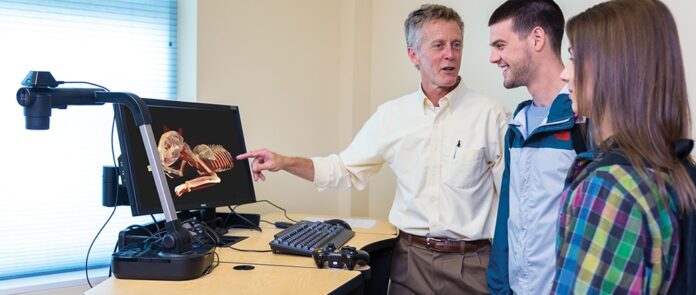
Virtual dissection or digital dissection provides students with the innovative learning opportunities in anatomy. Now, virtual dissection is achieved on anatomy visualization table. Patient CT scans are loaded on a near life-size computer screen and through the powerful software interactions, students can operate the data to accomplish their dissection. Importantly, virtually dissection of the CT scans, through touchscreen, students can work in groups to understand the complex anatomic relationships and one of the best techniques to prepare medical students for clinical practice (Darras et al.,13 2018; Darras et al.,45 2019). Unfortunately, students interacting with only touchscreen, they are not able to appreciate the way bone, tendons, muscle feel and loss of haptic feedback accompanies cadaveric dissection (Darras et al.,13 2018).
Social Media
Social media gained popularity in the anatomy field and becoming increasingly recognized as educational help to the anatomy educators (Pollock, Rea,46 2019). Facebook pages created by anatomists and most of the student users observing that interacting with the anatomy education pages helped their learning (Pickering, Bickerdike, 47 2017). The best way to teach and learn anatomy is through cadaveric dissection, but because the limited resources and access to such facilities so the social media could fill the gap (Iwanaga et al.,6 2020; Rai et al.,48 2019). Human cadavers or cadaveric material images or videos are being shared on public and this has a major ethical implication since there is no clarification of cadaveric source or consent received from the donors to share (Hennessy et al.,49 2020).
CONCLUSION
Traditional methods need to be replaced by the new technologies for veterinary anatomy teaching by using Plastination, Flash based modules, models, manikins and simulators which not only take care of animal welfare issues but also not let the purpose of learning to diminish. These approaches are cost effective and aids in better learning. Thus the use of new technologies in the field of anatomy education can be summarized by stating that the four R’s which comprise of Replacement, Reduction, Refinement and Rehabilitation should be used. Therefore,measures such as to minimize the number of animals used by altering the experimental design, reduction in the degree of experimental insult and substituting one species for another ( replacing higher animals from the lower animals) should be taken.
Compiled & Shared by- This paper is a compilation of groupwork provided by the Team, LITD (Livestock Institute of Training & Development)
Image-Courtesy-Google
Reference-On Request


