SURGICAL MANAGEMENT OF ABOMASAL FISTULA DUE TO GORE INJURY IN A MALE CALF – A CASE REPORT
- Vigneswari, M. Vidushi, S. Tina Roshini, N. Gurunathan, C. Chimithi, N. Arrivukkarasi and N. ArulJothi
Department of Veterinary Surgery and Radiology
Rajiv Gandhi Institute of Veterinary Education and Research, Puducherry
Abstract
A 4 months old CBJ male calf was presented to the Department of Veterinary Surgery and Radiology, VCC, RIVER, Puducherry with a history of gore injury on the umbilicus. On clinical examination, fistuluous tract with straw coloured fluid was noticed from the umbilicus. The pH of the fluid was acidic in nature and the case was confirmed as abomasal fistula. The animal was sedated with inj. Xylazine @ 0.1mg/kg b.wt I/V and inj. 2% lignocaine hydrochloride was infiltrated locally around the surgical site. Under aseptic condition, elliptical skin incision was made to resect the infected tissue and adhesions were separated between hernial sac and abomasum. The abomasal edges were debrided and sutured by cushing followed by lambert suture pattern using chromic catgut size 1. The abdominal cavity was lavaged with metronidazole solution. The hernial ring was closed using polyamide size 0 in overlapping suture pattern. Subcutaneous tissue and skin was closed routinely. Postoperatively inj. Streptopenicillin @ 10mg/kg, inj. Chlorpheniramine maleate @ 0.2mg/kg, inj. Meloxicam @ 0.2mg/kg for 5 days and 3 days, respectively. On 10th postoperative day sutures were removed and animal made an uneventful recovery.
Keywords: Abomasal fistula, calf
Introduction
Abomasal fistula is the continuous loss of digesta along with digestive secretions from an unusual site which may further alter the electrolyte and acid base status of animal (Dabas et al., 2016). It may be congenital or acquired. The present case describes about the surgical management of Abomasal fistula due to gore injury in a male calf.
Case history and observation
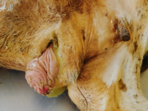
A 4 months old CBJ male calf was presented to the Department of Veterinary Surgery and Radiology, VCC, RIVER, Puducherry with a history of gore injury in the umbilicus. On clinical examination, fistuluous tract with straw coloured fluid was noticed from the umbilicus (Fig 1). The pH of the fluid was acidic in nature and the case was confirmed as abomasal fistula (Fig 2).
Surgical procedure
The animal was sedated with inj. Xylazine @ 0.1mg/kg b.wt I/V and inj. 2% lignocaine hydrochloride was infiltrated locally around the surgical site. Under aseptic condition, elliptical skin incision was made to resect the infected tissue and adhesions were separated between hernial sac and abomasum. The abomasal edges were debrided and sutured by cushing followed by lambert suture pattern using chromic catgut size 1 (Fig 3). The abdominal cavity was lavaged with metronidazole solution. The hernial ring was closed using polyamide size 0 in overlapping suture pattern. Subcutaneous tissue was sutured using catgut size 1 in walking suture pattern (Fig 4). Skin was opposed using polyamide size 0 in horizontal mattress suture pattern. Suture site was protected using benzoin seal and stent gauze applied. Postoperatively inj. Streptopenicillin @ 10mg/kg, inj. Chlorpheniramine maleate @ 0.2mg/kg, inj. Meloxicam @ 0.2mg/kg for 5 days and 3 days, respectively. On 10th postoperative day sutures were removed and animal made an uneventful recovery.
Discussion
Abomasal fistulation in association with ventral body wall hernias particularly umbilical hernias in calves have also been reported. Vertenten et al. (2009) has reported a case of abomasal fistula after a surgical procedure of displacement of abomasum in adult cattle, however abomasal fistula caused by gore injury in uncommon. In the present case, stab incision made by a local veterinarian on the swelling for the purpose of drainage seemed to be the most probable cause of fistula. The fistulation was found to be in the region of pylorus confirming the findings of Fubini and Ducharme (2004) and Sangwan et al. (2011). The present case report highlights the need of proper diagnosis followed by careful surgical intervention for treating the gore injury in calves.
Reference
- Dabas, V.S., Suthar, V.N and Chaudhari, C.F. (2016). Surgical Management of Umbilical Hernia with Abomasal Fistula in a Calf. Intas Polivet 17(I): 72-73.
- Fubini, S.L. and Ducharme, N.G. (2004). Surgery of abomasums. In:Farm Animal Surgery, (1st edn.). W.B. Saunders, New York, pp: 237.
- Sangwan, V., Kumar, A., Singh, K. and Mahajan, S.K. (2011). Umbilical Hernia with Abomasal- umbilical Fistula in a Cow Calf. Vet Scan. 6: 83.
4.Vertenten, G., Declercq, J., Gasthuys, F., Devisscher, L., Torfs, S., Van,L.G. and Martens, A. (2009). Abomasal end-to-end anastomosis as treatment for abomasal fistulation and herniation in a cow. Vet. Rec. 164: 785-786.
|
|
|
||
|
 |
||
|

|
||


