Know about Salter Harris Fracture : The fracture of the growing puppies and kittens
Definitions of “growth plate” and “Salter-Harris fracture”
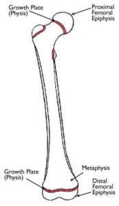
The growth plate also called “physis” is a thin line made of cartilage where the bone is growing in length in the immature growing animals (and humans). A Salter-Harris fracture is a fracture occurring over a growth plate which is a “weak line” of the bone structure. Hence, Salter-Harris fractures will only arise in animals younger than 11 months. During a trauma, the shock wave travels through the bone. Because the growth plate is a “weaker line”, the bone is more susceptible to break at this level and that defines the main mechanism and principles of a Salter-Harris fracture.
Where are growth plates located?
The growth plates are located at the long bones’ extremities; therefore, fractures at this level always occur nearby one of the following major joints: shoulder, elbow, wrist, hip, knee, and hock. Hence it is essential to properly treat those fractures.
How is a growth plate fracture suspected and diagnosed?
If your pet’s leg is fractured, this will necessarily cause a severe limp with heavy pain. Because fractures can be the result of a high-velocity trauma several lesions of the vital organs can concomitantly occur. Your veterinarian will perform a detailed and thorough physical examination and check that all vital signs are preserved. Often a blood test will be required as well.
What is the treatment for a Salter-Harris fracture?
As mentioned above, Salter-Harris fractures are happening at the level of a growth plate that is necessarily nearby joint. For this reason, it is imperative to treat these fractures at an early stage to preserve the continuity of the natural growth of the puppy/kitten as well as preserving the anatomy of the adjacent joint. Most Salter-Harris fractured should be treated with surgery and only a few of them can be treated conservatively with no surgery. Following surgery, an x-ray is performed to ensure proper positioning of the bone’s fragments and implants. A bandage is generally applied to reduce post-operative swelling and pain. In case no complication occurs, your pet can be discharged within 24 to 72 hours post-operatively.
The prognosis for a treated Salter-Harris fracture
Young animals are usually fragile, and their bones can break very easily; however, on the other hand, they also have a great healing potential thanks to developing and growing bone structure. If treated properly with surgery and early enough, a Salter-Harris fracture carries a “very good” to “excellent” prognosis. Rarely on ossification of the fractured growth plate will occur causing the bone to curve or stunting the growth. Several weeks after the initial surgery, physiotherapy might be required for a better functional recovery of the operated leg.
Salter-Harris Classification

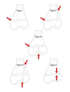
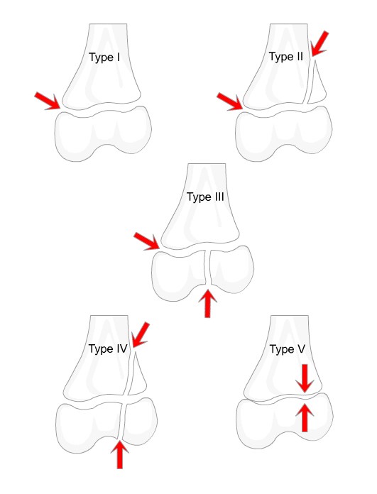
This physeal fracture classification system was first described in 1963 by Salter and Harris .
Type I
Type I – transverse fracture through the physis. The epiphysis separates completely from the metaphysis.
Type II
Type II – fracture through the physis with a detached triangular metaphyseal fragment. This is the most common type of physeal injury seen.
Type III
Type III – a fracture through the physis and then entering the joint through a fracture of the epiphysis. This is therefore an intra-articular fracture but it is very rare.
Type IV
Type IV – a fracture through the epiphysis, physis and metaphysis involving all three areas and being again intra-articular.
Type V
Type V – crush injury of the physis. There may be very little evidence of this on initial x-ray but the damaged physis means abnormal appearances months or years later as the bone growth has arrested.
S.A.L.T.R
To help remember these different types use the S.A.L.T.R mnemonic:
1 = Slip
2 = Above the physis (through metaphysis)
3 = Lower than the physis (through epiphysis)
4 = Through (metaphysis, growth plate and epiphysis)
5 = Ram (a crushing type injury)

Compiled & Shared by- This paper is a compilation of groupwork provided by the Team, LITD (Livestock Institute of Training & Development)
Image-Courtesy-Google
Reference-On Request


