


BLOOD TRANSFUSION IN ANIMALS
Shivani Bante1, Vivek Kumar Maurya2, Jagmohan Rajput2 and Anushka Gupta3
- VAS, IAHVB, Mhow, Govt. of MP
- MVSc. scholar, College of Veterinary Science & A.H., Mhow.
- BVSc & AH scholar, College of Veterinary Science & A.H., Mhow
INTRODUCTION
Blood transfusion is being practiced for centuries for saving life of human beings and animals. Richard Lower in 1665 transfused the blood in a dog for the first time in the history. With the help of latest techniques and equipment developed after 1950, blood transfusion became more popular in veterinary medicine. Blood transfusion has made considerable advancements in veterinary medicine in recent times. Although, the information and availability of blood and its products has increased, transfusion therapy has become more complex. Advanced screening facilities, blood group testing and techniques for cross matching blood had made the process of donor selection more complicated. Advancement in techniques of separating the components of blood has given the clinician an opportunity to use the component as per the demand of the patient.
Farm animals especially in bovine, severe anemia occurs due to blood protozoan infection like Theileriosis, Babesiosis, Anaplasmosis, ecto and endo parasite, blood loss during surgery and nutritional deficiency. In severe anemia where there is extreme depletion of oxygen carrying capacity of the blood occurs, Most of the animals die due to severe anemia. Therefore, a simple, rapid, inexpensive and timely administration of blood in cases of life-threatening anemia in large animal is clinically very rewarding. By keeping in view of all those things Blood transfusion kit for blood transfusion in large animals is designed. Blood transfusion kit is easy to perform, less time consuming, economically justifiable, giving spectacular results and professional.
INDICATIONS FOR BLOOD TRANSFUSIONS:
- Acute haemorrhage
-Complication after caesarian section.
-Bleeding due to uterine prolapse.
-Umbilical vessel damage by a new born calf.
-Accidental Trauma.
-Post parturient hemoglobinurea.
-Hypoposphatemia in buffalo. - Blood Protozoan infection
-Theileriosis.
-Babesiosis.
-Anaplasmosis. - Abomasal ulceration.
- Anemia and melena.
(Animal with severe anaemia (<10% PCV) – These cases are likely to die regardless of other treatments.)
(Animal with moderate anaemia (10-15% PCV) – A transfusion is recommended to aid in recovery.)
BLOOD GROUPS
Blood groups are named according to the species-specific antigens present on the surface of erythrocytes. Theses antigens play an important role in inducing immune-mediated reactions and can cause complications while transfusing blood from different blood groups. Antigens coupled with platelets, leukocytes and plasma proteins may also induce immune mediated reactions in host animals during transfusion therapies. Plasma also has some naturally occurring all antibodies that can act against other blood groups without any prior exposure to the erythrocyte antigens. Erythrocyte antigens can induce production of antibodies when animals get exposed via blood transfusion, transplacental exposure or in the case of neonatal isoerythrolysis (NI), through colostrum. Blood groups in the common domestic and pet animal species are described here. From a clinician’s point of view, these are the antigens to which the veterinary practitioner should be most familiar. However, many other blood group factors and systems have been described and the lack of commercially available typing sera does not diminish the potential significance of these other systems in transfusion medicine.
Dog
International workshops met in 1972 and 1974 to standardize canine blood groups as defined by isoimmune sera and to standardize canine blood group system nomenclature. The first workshop designated the terminology canine erythrocyte antigen (CEA) followed by a number to indicate the blood group antigen. The second workshop adopted the designation dog erythrocyte antigen (DEA). The new terminology was adopted to avoid confusion with the carcinoembryonic antigen (CEA) system. The blood group system in dogs includes DEA 1.1, DEA 1.2, DEA 3, DEA 4, DEA 5 and DEA 7. The DEA nomenclature system of canine blood group is not accepted worldwide, although some authors use the newer genetic nomenclature system in reporting new blood group specificities (Symons and Bell, 1992). In dogs, naturally occurring alloantibodies are of lesser clinical significance whereas in cats it is very important clinically. DEA 1.1 and 1.2 are the most important blood groups and are found in 60% population of canines. These blood groups can elicit severe transfusion reactions in previously sensitized dogs.
 |
|
|
|
DEA 1.3 has been described in German shepherd dogs in Australia. DEA 4 blood group of dogs is found in high frequency that can cause hemolytic transfusion reactions in DEA 4-negative dogs previously sensitized by DEA 4-positive blood transfusions. The DEA 3, 5 and 7 blood groups can cause delayed transfusion reactions in dogs lacking these antigens but are previously sensitized to these antigen
Cat
Three blood groups are reported in cats in AB blood group system. Type A blood group is the most common group and found in 95% of the American cats. Majority of Indian and 30% of cats in UK belongs to blood group B. Blood group AB is extremely rare but is found in DSH/DLH cats and in breeds in which group B is also found e.g. Abyssinian, Birman, British shorthair, Norwegian forest, Somali, Scottish fold and Persian. One more erythrocyte antigen, a novel Mik antigen, has been reported in DSH cats. One should consider the variation in geographical variation in blood groups of felids. The risk of transfusing lethal groups A or AB to a cat of blood group B is 20%. Anti-A, anti-B and anti-Mik are naturally occurring alloantibodies found in cats. Alloantibodies that are strong hemagglutinins and hemolysins against type A erythrocytes are very rich in serum of cats with blood group B. Type A cats generally have weak hemagglutinins and hemolysins against type B erythrocytes, hence transfusion reactions are rare. Neonatal isoerythrolysis (NI) occurs in type A or AB suckling kittens born to type B queens with transfer of the anti-A alloantibodies via the colostral transfer of immunoglobulin (primarily IgG). Transfusion between Mik positive and Mik negative cats can result in acute post-transfusion hemolysins.
Horse and donkey
The seven blood groups in horses viz. A, C, D, K, P, Q and U are internationally recognized with more than 30 erythrocyte antigens. Universal donor horse is not possible because of various possible antigenic combinations. The cross matching must be performed although impractical to minimize transfusion reactions. Aa and Qa alloantigens are hemolysins and are extremely immunogenic and most cases of NI are associated with anti-Aa or -Qa antibodies. In horses Blood group vary with breeds. Thoroughbreds and Arabian breeds have high prevalence of antigens Aa or Qa whereas, Standard breds lack the Qa antigen. Donkey factor, a unique donkey and mule erythrocyte antigen is not found in the horse and is responsible for neonatal isoerythrolysis in mule pregnancies.
Cattle
The internationally recognized blood groups in cattle are A, B, C, F, J, L, M, R, S, T and Z. out of these 11 groups, group B and J being the most clinically relevant. The B group itself has more than 60 antigens, thereby making closely matched blood transfusions difficult. The J antigen is not a true erythrocyte antigen but a lipid found in plasma Cattle having anti-J antibodies with a small amount of adsorbed J antigen on erythrocytes but negative J blood group, can develop transfusion reactions when receiving J-positive blood.
Sheep and goat
A, B, C, D, M, R, X are the seven blood groups identified in sheep. The B group has over 52 factors present over erythrocytes. The R system in sheep is similar to the J system in cattle (i.e., antigens are soluble and passively adsorbed to erythrocytes). The M-L blood group in sheep is related to active potassium transport in reticulocytes. The blood groups of the goat (A, B, C, M, J) are very similar to those of sheep with the B system equally complex. Many of the reagents used for blood typing of sheep also have been used to type goats.
GENERAL PRINCIPLES OF CROSS MATCHING BLOOD
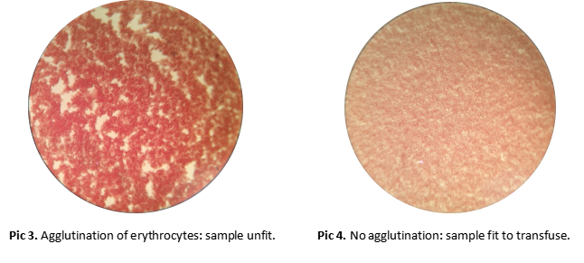
Major and minor cross matching tests are done for agglutinating and/or hemolytic reactions between donor and recipient. For dog and cat agglutinating tests are sufficient whereas in equines agglutinating and hemolytic tests are required because of presence of agglutinating as well as hemolytic antibodies in equines. For cattle, sheep and goats test for hemolytic antibodies and complement is necessary. The major cross match evaluates for the presence (positive findings) or absence (negative findings) of detectable levels of antibodies, whether naturally occurring or induced, in the recipient against donor erythrocyte antigens. A major cross match should always be performed in animals that have strong naturally occurring antibodies, as in cats, or in those that may have induced antibodies as from prior transfusions. The latter is true even if the same donor blood is intended for repeated transfusion beyond a span of several days. The minor cross match procedure follows the same steps as the major cross match but evaluates for the presence or absence of detectable antibodies in donor plasma against recipient erythrocytes. Minor cross matching is of little significance because the volume of plasma donated is very small in comparison to the recipient and is diluted in the recipient particularly in case when only erythrocytes are transfused. Administration of packed erythrocytes may contain sufficient antibodies against recipient erythrocytes to induce adverse reactions in dogs and horses. An ethylene diamine tetraacetic acid (EDTA) tube and a clot tube from the recipient are preferred for use in cross match testing. The EDTA plasma should not be used in place of serum because this contributes to increased rouleaux formation and difficult interpretation of agglutination, particularly in the horse. Preferably, samples should be free of auto-agglutination, hemolysis and lipemia to aid in the interpretation of the reactions. When auto agglutination is present, or when no compatible units are available, transfusing the least incompatible unit may be a necessity, albeit not without significant risk. Test transfusing even a small volume of unmatched blood is an unsafe practice and never recommended.
Transfusion Reaction Signs:
- Increase of respiratory rate
• Urticaria
• Hiccupping
• Sweating
• Tachycardia
• Violent movements
• Severe respiratory distress
CONCLUSION
Transfusion medicine may be a lifesaving modality in case of emergency or critically ill animals. Blood products are becoming readily available and transfusions can be performed in many veterinary clinics. The appropriate use of transfusion medicine should balance the rare but not negligible potential risks associated with transfusions. Patients should be appropriately screened with blood typing and cross matching before transfusion. Ongoing research may provide even better platform typing, longer storage times and a larger variety of products that are more specific for each species. Blood transfusion kit is very helpful for successful blood transfusion in large animals because all the material, chemicals, medicine and procedure are given in kit.