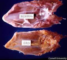
CHRONIC RESPIRATORY DISEASE (CRD) IN POULTRY
The disease has been reported in chickens and turkeys. CRD is specific disease caused by one of the group of organisms known is pleuro pneumonia like organism (PPLO), but more closely defined is Mycoplasma; the particular organism directly associated with CRD is Alycoplasma gallisepticum with or without any secondary complications. According to I he recommendation of FAQ committee meeting held in May l969, the term “Avian Respiratory Mycoplasmosis” (ARM) be used in uncomplicated outbreaks involving only pathogenic avian PPLC (Mycoplasma) and the term CRD be used when PPLO infection is superimposed with other condition
The mortality entirely to CRD is negligible, but it is important because it predisposes the birds to infection for other disease producing organisms.
Transmission:
M. gallisepticum is transmitted through eggs but organisms can also pass from bird to bird through nasal discharges and through droppings. It can also be transmitted by hands, feet and clothes of attendants of visitors. Symptoms: Uncomplicated CRD is frequently sub-clinical. When symptoms are present they are normally milk in nature and include coughing, sneezing, and a nasal discharge. In turkeys, sinuses are frequently swollen. On postmortem examination the trachea may be found inflamed and the air sacs thickened with pus. The condition affecting air sac is often referred as “air sac” disease but it is more pronounced when other factors including bacteria, complicate the original CRD infection.
The- organism has long incubation period of 10 to 30 days. Therefore only few outbreaks-seen in birds under 4 weeks old
Mycoplasma gallisepticum is considered the primary cause of Chronic Respiratory Disease (CRD) in poultry. The most common symptoms of Mycoplasma gallisepticum infections in poultry are eye problems and inflammation around the face and cere. Other symptoms include open mouth breathing and gurgling throat sounds.
the eye), pussy eye discharge, sticky eyelids and open mouth breathing. Afflicted birds and the entire flock should be treated when this stage of the disease is seen. Treatment involves the administration of a combination of antibiotics (e.g. doxycycline hydrochloride and tylosine tartrate) into the drinking water for 7 days. An appropriate eye cream is applied to those birds with eye symptoms for 2 days. To accelerate recovery and help reduce the effects of any stressful factors, Turbobooster and E-powder should be mixed into a seed treat each day for 7 days. For stage two of this disease this treatment should give a good response. A poor response indicates that underlying stress factors remain and if not seen to, the disease will progress to stage three.
Mode of transmission
The disease is spread both vertically and horizontally.
Vertical Transmission
The organism can be incorporated into eggs by infected breeders and chickens hatched carrying the mycoplasma infection.
Horizontal Transmission
Disease transmission may also take place from direct contact with infected birds and will spread throughout the flock in this way. Transmission may also occur by contact of healthy birds with equipments contaminated by infected birds.
It is usually transmitted through eggs, feed, water, droplet infection, and by care takers (5). E. coli is a secondary invader of Mycoplasma-infected birds. Aerosol spread occurs. Severe ND, IBV or ILT reactions may occur after vaccination, especially if given by spray to chicks previously infected with Mycoplasma or E. coli.
Predisposing factors
Environmental stress factors such as increased or decreased temperature, high concentration of ammonia, dust in the air, and other factors associated with poor ventilation, may act as predisposing factors.
Clinical signs (Fig 1&2)
The disease is characterized by loss of appetite, dullness, depression, tendency to huddle together, coughing, tail bobbing when breathing, emaciation, respiratory rales, sneezing, open mouth breathing, conjunctivitis, periorbital swelling, poor growth, decreased feed consumption and increase in mortality (6 & 7).
In Layers / Breeders
• Nasal and ocular discharge, (watery eyes) rattling in the wind pipes, coughing, gasping (dyspnea), sneezing and shaking of the hed.
• Feed consumption drops off leading to decreased egg production and loss of weight.
• Male birds frequently have the most prominent signs.
• Reduced hatchability and chick viability.
• Occasional encephalopathy and abnormal feathers
In Broilers
• Most outbreaks occur between 3rd and 6th weeks of age .
• Poor feed conversion, sharp decline in weight gain.
• Slow growth
• Leg problems
• Morbidity rate fairly high but not great mortality.
• Poor carcass quality, high contamination rate. Thin and weak birds with razor-blade breasts.
In Turkeys
• often accompanied by swelling of the paranasal (infraorbital) sinus.
• Conjunctivitis with a frothy ocular exudate
• Production is lower in infected flocks, decreased weight gain, feed efficiency and egg production.
Fig. 1 Poultry showing dullness and depression (8)
Fig. 2 Poultry showing conjunctivitis, periorbital swelling and eyelid edema (8)
Postmortem lesions (Fig 3-12)
• Airsacculitis
• Fibrinous pericarditis and perihepatitis
• Salpingitis
• Panopthalmitis
• Presence of caseous exudates in the air sac
• Severe tracheitis with variable exudate – catarrhal to purulent
• Pulmonary granuloma (6, 9 & 10)
Fig. 3 Cloudy appearance of the abdominal airsacs (11)
Fig. 4 Pericarditis and perihepatitis (11)
Fig. 5 Severe tracheitis. Both tracheas are congested and the upper sample has mucopurulent material in the lumen (12)
Fig. 6 Air sac showing coccobacilliform bodies surrounded by mononuclear cells (13)
Fig. 7 Air sac showing presence of fibrinous and serofibrinous exudates (13)
Fig. 8 Lung showing accumulation of serous exudates in tertiary bronchioles and atria. Increased cellularity of air capillaries (14)
Fig. 9 Giant cell granuloma in air capillaries (14)
Fig. 10 Accumulation of haemorrhagic exudates in tertiary bronchioles and atria (14)
Fig.11 Larynx showing lymphofollicular reaction in submucosa (14)
Fig. 12 Trachea showing hypertrophy, hydropic degeneration and tuboalveolar elongation of intraepithelial glands (14)
Diagnosis:
• Laboratory isolation and identification of E. coli or Mycoplasma from lesions (Fig 13-16).
• Detection of Mycoplasma colonies using fluorescent antibody test, recombinant probe and hybridisation or antigen capture ELISA.
Biochemical studies (15)
• Tetrazolium methylene blue reduction test
• Carbohydrate fermantation test
• Breakdown of arginine
• Formation of film on egg yolk agar
Serological test (16 & 17)
• Growth inhibition test
• ELISA
• Plate agglutination
• Haemagglutination-inhibition, (HI)
• Testing of sera for antibodies against Mycoplasma.
Molecular techniques (18)
• Polymerase chain reaction
Fig. 13 & 14 Mycoplasmal colonies on Edward’s PPLO agar (19)
Fig. 15 & 16 Mycoplasmal colonies with Diene’s stain on Edward’s PPLO agar (19)
Prevention and control:
As uncomplicated CRD is not a major problem today, it may be necessary to protect only against the probable complicating factors. Increased ventilation without drafts reduces the spread and severity of CRD. Buying replacement stock from CRD free source greatly reduces the risk of spread. A very high standard of hygienic condition is of course, supremely important.
It is difficult to prevent infections caused by Mycoplasma gallisepticum because the disease is transmitted by egg and any new birds must be free of the disease. Vaccinations have not proven to be a successful preventative measure because CRD is so often complicated by underlying diseases. Careful management strategies that minimise stress and the ability to determine the stage of CRD are important preventative measures in the treatment and long term prevention of Mycoplasma gallisepticum infections.
The basic control measures suggested are–
• Proper biosecurity program and schedule, for each cycle
Establishment of Mycoplasma free breeding flocks.
Stock free of Mycoplasma infection can be produced by vaccination. Vaccinate breeders against E. coli, MG, MS, NDV, IBV, ILT and infectious bursal disease (IBD) to prevent the disease.
Vaccinate progeny against ND, IB, ILT and IBD, hatch and place Mycoplasma infected stock separate from mycoplasma negative stock to reduce the spread of the organism. Treat all Mycoplasma positive flocks with antibiotics to reduce spread into eggs.
• Treating infected hatching eggs with the antibiotic to kill the organism contained in the eggs.
• Before purchasing chicks from a hatchery, it should be confirmed that they are free from CRD.
• Chicks should be raised at the place where there is no approach of infected birds.
• Complete fencing of the breeding farms and sufficient isolation of prevent airborne infections from infected flocks.
• Disposing of dead birds by incineration, deep burial or by means of special disposal pits.
• Using vaccines that are free from contamination of Mycoplasma gallisepticum.
• Construction of the houses must be done in such a way that prohibit the entrance of any type of wild birds and wandering animals.
• Prohibition of visitors in the farm.
• Before coming in contact with flocks, workmen should take shower and put on special clothes.
• Strict bio-security measures should be adopted.
Treatment:
The tetracycline group of drugs is useful in treatment, if given continuously for over a week, as soon as disease’s seen in flock, at the rate of 100-400 g per ton of feed. It can also be given through water. Nitrofurans especially furazalidone is very effective. Streptomycin may be injected in sinuses after removal of mucous by spraying in turkeys are helpful.
Disinfect the farm and equipments with right disinfectant.
• Mycoplasmas are resistant to antibiotics that act on cell wall, such as penicillin, but are sensitive to tetracyclines (oxytetracycline, chlortetracycline and doxycycline), macrolides (erythromycin, tylosin, spiramycin, lincomycin, and kitasamycin), quinolones (imequil, norfloxacin, enrofloxacin and levofloxacin) or tiamulin. Drugs that accumulate in high concentrations in the mucosal membranes of the respiratory and genitourinary tracts, such as tiamulin and quinolones( enrofloxacin, specially levofloxacin) are recommended.
For CCRD
• If the mycoplasmosis is clubbed with other bacterial infections like E.coli administer mycoplasmosis drugs with gram -ve levofloxacin with colistin or Neomycin and Doxycycline through drinking water in addition to the above treatment for 3 to 5 days.
For Chicks
• Chicks arrived from known infected parent flocks should be treated with a suitable antibiotic( levofloxacin with colistin) during the first 48 hours after placement and then subsequently at 20 – 24 days for 24 to 48 hours period.
• Efforts should be made to reduce dust and secondary infections. Improve the ventilation for having good results of medicine.
• Medicate all Mycoplasma-positive broilers for the first 7-10 days in the feed or water.
• Ethereal oils based Herbal bronchodialator and anti-inflammatory agents are increasingly becoming acceptable to veterinarians, because they are free from side effects compared to allopathic contemporaries and deliver quicker symptomatic relief compared to allopathic as well as herbal (cough syrups) contemporaries.
Therapy of CCRD is complex, and there is really no sure shot treatment to completely eliminate Mycoplasma- infected birds, except elimination of the flock.
Fluoroquinolones (Enrofloxacin, Danofloxacin, Norfloxacin, Flumequin etc.) are on withdrawal phase world- wide, owing to their deleterious effects to public health through cross resistance to human strains of Campylobacter and other bacteria.
Various antibiotics like Tylosin, Tiamulin, Tilmicosin, Aivlosin, Tetracyclines (mainly Doxycycline, Chlortetracycline, Oxytetracycline), Spiramycin, Erythromycin, Gentamicin and Ketasamycin, Neomycin, Colistin, etc. are being used alone or in various combinations to cure and control the disease, with variable success, owing to mixed response to resistance development in the various microbes
A preventive program against CCRD has been developed and tested successfully by Montajat, which alleviates any passive infection. In this program, following regimen is followed during the broiler life- cycle:
- Day 1, 2 ,3 oral liquid of Tilmicosin 25% @ 1ml/ 3 liters of drinking water, along with Colistin sulphate 10MIU/gm @ 1gm/ 10 liters of drinking water. It cleans the chicks from Mycoplasma sp., E. coli and Salmonella brought in the flock vertically or horizontally, or vaccine reaction flare.
- Day 13, 14 and 15 (or equivalent for 3 days, after one day of Gumboro vaccination). Administer Florfenicol 30% oral solution @ 30mg per kg body weight (grossly 1 liter/ 10MT of live bodyweight), to clear all E. coli and Salmonella sp.
HOMEOPATHIC TREATMENT–
Combination of Thuja200+Nat sulf200+Carboveg200+Bryonia200, 5ml each in 8 liter of water for 100 birds for 3 days.
Compiled  & Shared by- Team, LITD (Livestock Institute of Training & Development)


Image-Courtesy-Google


Reference-On Request.


