A Brief Introduction to Gangrenous Dermatitis in Poultry
Neetesh kumar 1, Akshit Tyagi 2, Dharam Prkash Srivastava3*, Anwesha Baag2, Surya Kant4, Himanshu Gautum5
- PhD Scholar, Department of Veterinary Pathology
- PG Scholar, Department of Veterinary Pathology
- SMS, KVK PG College Ghazipur
- PG Scholar, Department of Livestock Production and Management
- PG Scholar, Department of Veterinary Medicine
College of Veterinary Science and Animal Husbandry A.N.D.U.A.T., Kumarganj
Ayodhya, U.P. -224229
Corresponding Author: *dr.dpsrivastava56@gmail.com
Introduction
Gangrenous dermatitis (GD) is a disease that mainly affects fast growing broiler chickens of age between 4 to 8 weeks and causes severe economical loss to poultry industry worldwide. The disease is also known by other names like necrotic dermatitis, gangrenous cellulitis, gangrenous dermatomyositis, poultry gangrene, avian malignant edema, gas edema disease, subcutaneous emphysema, blue wing disease, and wing rot.
The disease is characterized by congestion, hemorrhage, and necrosis of the skin and subcutaneous tissue, associated with edema or emphysema, which sometimes extends into the underlying musculature. The affected areas mostly include breast, back, abdomen, thighs, tail and wings of chicks.
Etiology
A wound to the skin usually starts disease which is quickly followed by a secondary bacterial infection.Secondary bacterial infection is mainly caused by Clostridium perfringens type A, Clostridium septicum or Staphylococcus aureus.
Genus Clostridium, is composed of anaerobic, mostly gram-positive, spore-forming rods whereas Genus Staphylococcus is composed of gram-positive, coccoid-shaped, aerobic bacteria, which are commonly seen as clusters when grown in solid media and short chains when cultured in liquid media.
- perfringenstype A isolates produce alpha toxin (CPA); some strains may also produce one or more additional toxins including necrotic enteritis B–like toxin (NetB) and enterotoxin (CPE).
The main virulence factor of C. septicum is alpha toxin (ATX), a necrotizing pore-forming toxin (PFT), which is unrelated to the alpha toxin of C. perfringens. ATX induces increased membrane permeability, which leads to cell necrosis. S. aureus can produce several toxins, including hyaluronidase, deoxyribonuclease, fibrinolysin, lipase, protease, leucocidin, hemolysins, epidermolytic toxin, and dermonecrotic toxin.
These bacteria can act alone or along with combination of different anaerobic and aerobic bacteria including Clostridium sordellii, Clostridium novyi, Staphylococcus aureus, Staphylococcus epidermidis, Staphylococcus xylosus, Escherichia coli, Enterococcus faecalis, Pasteurella multocida, Gallibacterium anatis biovar haemolytica, Proteus spp., Pseudomonas aeruginosa, Bacillus spp., and Erysipelothrix rhusiopathiae .
These bacteria are usually not able to penetrate intact skin. They infectious agents can be ingested if healthy birds peck at dead birds that have died with the disease or if litter and feces are contaminated with large numbers of disease-causing organisms
Pathogenesis
Pathogenesis of disease is due to many predisposing factors in which immunosuppression may be the key predisposing factor for GD in chickens. Under natural conditions, immunosuppression can be triggered by a wide range of infectious and environmental factors in chickens.
Immunosuppressive viral agents that may predispose to GD in chicken include Marek’s disease virus, infectious bursal disease virus, chicken anemia virus, several reoviruses, and inclusion body hepatitis virus in chickens
Environmental factors that can predispose chickens to GD are traumatic lesions of the skin associated with cannibalism or fighting, Overcrowding, feed outages, deficient diets, wet and poor litter conditions, contaminated feed, water, equipment, and vaccines, high ammonia levels and mycotoxins in feed
In broiler chickens, GD is mainly predisposed to by traumatic damage of the skin, usually associated with cannibalism and overcrowding. Such skin lesions provide a port of entry for bacteria. Chaotic multiplication of the intestinal flora followed by absorption into the bloodstream promoted bacteremia, which is also the origin for some of the GD lesions. Outbreaks seem to be greater in summer and fall than in winter and spring
Clinical signs
The disease can occur without clinical signs being observed. However, high fever, depression, anorexia, ataxia, leg weakness, and lateral recumbency are usually seen. The lower abdomen and inner thighs are frequently affected by the accumulation of subcutaneous edema.
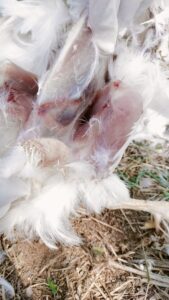
Fig 1: Gangrenous Dermatitis in birds
The skin over affected areas is usually featherless and can show dark-red, purple, green, or green-blue discoloration. The most frequently affected areas of the body are breast, abdomen, back, thighs, legs, and wings of chicken.
Lesions
Microscopic lesions are characterized by oedema, emphysema, hyperaemia, haemorrhages and necroses in the subcutaneous tissues.
Gross Lesions
A predominant feature of GD is rapid autolysis, which is more prevalent in cases of sudden death. Severe edema mixed with gas and hemorrhages in the subcutaneous tissue are present. Abrasions are frequently found on the skin. The muscle under skin lesions may show gray or tan discoloration, hemorrhages, edema, and emphysema. The feathers can be easily removed from the affected skin areas.
DIAGNOSIS
Presumptive diagnosis of GD can be based on clinical signs (fever, listlessness, anorexia, ataxia, and recumbency), Microscopic finding, Detection of bacteria, Gross findings, Epidemiology and post mortem changes
Detection of bacteria can be achieved by aerobic and anaerobic culture, fluorescent antibodies, immunohistochemistry, and PCR assay
Microscopic findings like gram-positive rods on smears of the serosanguineous exudate, collected from affected skin or subcutaneous tissue, adds certainty to the presumptive diagnosis
Confirmation should rely on detection of the agents involved, associated with the clinical signs and gross and microscopic findings. It is important to remember that isolation of any of the agents of GD without clinical signs and/or lesions of the disease is not diagnostic
Prevention
Following are key management strategies that poultry producers can implement to help with disease prevention
- Ensure a robust immune status.
- Reduce the level of clostridium in the environment.
- Minimize injury to the skin and intestinal tract, which are the two main route of infection.
To help reduce immunosuppression in poultry flocks,proper vaccination against Infectious bursal disease, Marek’s, chick anemia virus and reovirus is important for protecting the broiler’s immune system. An environment that promotes poor litter conditions may also predispose flocks to GD. Good litter management, immediately addressing water leaks, and regular pickup and proper disposal of affected dead multiple times a day are necessary to keep barn clostridium at low levels. Affected poultry farms are likely to have repeat outbreaks if the environment if not treated properly. It is recommended to do a full litter clean-out between flocks along with reduced stocking density. Treatment of the barn floor with a product that can lower the pH can reduce repetitive breaks on subsequent flocks.
Intestinal Integrity provided by an effective anticoccidial program, as well as pro and prebiotics, helps maintain a healthy intestinal micro flora and a tight intestinal barrier that keeps clostridium or other disease-causing bacteria found in the intestine from passing into the blood and causing infection. Products can help get the flora found in the intestinal tract back in balance and may help reduce incidence and mortality. However, since Clostridial is critical to the disease, a healthy microflora does not always reduce disease.
Growers should closely monitor feeding status and shedule. Do not let your birds run out of feed. Bird activity increases when hungry chickens receive feed, resulting in an increased number of cuts, scratches, and skin damage. Also, be sure that migration fences are in place on time to prevent overcrowding in some areas of the house that may increase the GD risk. Avoid loud noises that may disturb birds and increase likelihood of cuts, scratches, and skin damage. Follow a lighting program that helps calm the birds and control activity level. A proper management wil reduce the stress for the birds and reduces the possibility of a skin injury will reduce the risk of a GD outbreak.
Treatment
Antibiotics in starter feed to reduce bacteria
Effective antibiotics against gangrenous dermatitis include penicillin, erythromycin and tetracycline group of antibiotics
Post Mortem Findings
Post mortem findings usually include gross findings like skin and subcutaneous necrosis, edema, emphysema, and hemorrhage. Microscopic findings like necrotizing dermatitis and cellulitis with hemorrhage and emphysema is also seen in during post mortem.
Refrences
https://www.ncbi.nlm.nih.gov/pmc/articles/PMC6505868/#bibr42-1040638717742435


