


Application of Exfoliative Vaginal Cytology (EVC) as a Tool for Determining Breeding Time in Dogs & Cats
Vaginal cytology is an easy and cheap method with great value in small animal reproduction. As a general introduction in function of cellular types that we can see, we can determine the phase of the cycle in the bitch or queen. First we will describe the types of cells that we have and then the correlation with the ovarian cycle. The alterations in % and types of cells are directly related to serum estrogen concentration. Estrogens are the hormones secreted by a maturing follicles and cause a thickening of vaginal linin, moving the cells from her blood supply, with the subsequent degeneration and atrophy. Thus, vaginal cytology is always a reflex of estrogen activity on the female.
Techniques in vaginal smears
The preferred method to obtain a good vaginal cytology, is the application of a cotton swab in the cranial vagina of a bitch . The lip of the vulva are gently separated with the fingers and a cotton swab is passed into the dorsal aspect of the vulva. We must avoid clitoris fossa and advance in a craniodorsal direction, toward vertebral column. At this point we rotate the cotton swab and withdraw it. We must observe the color, and type of secretions and rotate gently the cotton tip in a slide. Usually three or four slides are fixed every time, with the name and date. A lot of stains has been described, but usually Diff-Quick, new methylene blue and Schor’s stain modification (with criteria for keratinization) are used.
In the queen the cotton swab must be of adequate size (urethral human culturette tube) and introduced only 1 to 1,5 cm in the vagina. The main differences between queen and bitch is the relatively straight horizontal anatomy of the vestibule of the vagina, the vagina, and the cervix. Usually when we introduce the cotton swab we are inducing the ovulation of queen (cervico-hypothalamus reflex) with the subsequent external behaviour aspect (vocalization and others).
Examination of exfoliated cells from the vagina is a simple technique that is useful to monitor the progression of proestrus and estrus in dogs and cats.The vaginal epithelium undergoes a predictable hyperplastic response to increasing plasma estrogen concentrations during proestrus. Starting as only a few cell layers, the epithelium becomes 20 to 30 cell layers thick, eventually exfoliating large numbers of superficial epithelial cells during estrus. Vaginal cytology, often in tandem with hormone analysis, may provide valuable information about the stage of the ovarian cycle.In addition, vaginal cytology has proven useful to detect inflammatory and neoplastic conditions in the female reproductive tract.
Performing vaginal cytology offers a rapid, inexpensive, and reliable in-clinic method to evaluate stages of the estrus cycle in the bitch. Veterinary discomfort with obtaining and interpreting vaginal cytology is common; submission to a commercial laboratory might result in diagnostic delays and increased client costs Equipment required for vaginal cytology (cotton-tipped applicators, frosted microscope slides, commercial Romanowsky [Diff-Quik] stain, and light microscope) is already present in most small animal practices.
Competence in vaginal cytology allows a clinician to:
*Determine whether a bitch is actually in heat.
*Aid in determining the correct time to begin performing more expensive serum progesterone and luteinizing hormone (LH) assays for precise ovulation timing.
*Determine if it is too late in the estrus cycle to perform artificial insemination in dogs unable or unwilling to breed naturally.
*Determine if a bitch is under the influence of estrogen (endogenous or exogenous).
*Predict the correct day to perform an elective cesarean section (C-section)
Collecting Vaginal Samples
Cells are obtained by passing a cotton-tipped swab into the caudal vagina . A narrow spreading speculum may be used to allow unimpeded swab passage. If no vaginal discharge is present, the swab may be moistened with sterile saline to avoid discomfort. The swab should be directed craniodorsad when entering the vaginal vault to avoid the clitoral fossa; keratinized epithelium normally present in the fossa could lead to an inappropriate cytologic interpretation .Once cranial to the urethral orifice, the vaginal wall is gently swabbed. The cells are then transferred to a glass slide by gently rolling the swab with minimal pressure to avoid rupturing cells. The smear is allowed to air-dry thoroughly before staining. Romanowsky-type stains typically used for blood films (Wright or modified Wright-Giemsa stains, including quick-type stains) provide good morphologic detail.

The vaginal epithelium is responsive to sex steroids, particularly estrogen, and undergoes predictable changes through the cycle in response to changes in blood concentrations of ovarian hormones. Rising levels of estrogen cause the vaginal epithelium to become “cornified” – the surface cells become large and flattened, with small or absent nuclei.
In essence, vaginal cytology is a type of endocrine assay. Tracking changes in the morphology of desquamated vaginal epithelial cells provides a convenient means of assaying changes in estrogen levels. The technique is used widely in managing canine breeding programs.
Techniques for Preparing a Canine Vaginal Smear
Materials Required to Obtain and Process Vaginal Smears
- cotton-tipped swabs (six inch variety)
- microscope slides (have these close by where the smear will be taken)
- methanol or commercial spray fixative for cytology (can also use ethanol)
- Diff-Quik staining solution (American Scientific Products) or a stain such as Giemsa or Wrights that is used for staining blood smears
Obtaining the Sample
The objective is to obtain a sample of epithelial cells from the vagina, and one should avoid sampling from the vestibule (i.e. just inside the vulva). Even in small dogs, the swab should be inserted several inches past the vulva; in large breed dogs (e.g. German Shepard bitch), a majority of the 6-inch swab can be inserted. Bitches in proestrus or estrus rarely object to this procedure, although some restraint may be required to prevent “squirming”.
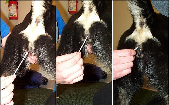
Part the lips of the vulva and gently insert a swab at a relatively steep angle. Some people suggest using a speculum (e.g. the cone from an otoscope) and some like to moisten the swab with saline prior to use, but neither of this is critical. After 1-2 inches of the swab have been inserted, the angle of insertion can be altered to roughly 45 degrees and insertion continued.
When the swab is fully inserted, rotate the end through 2-3 revolutions, which will allow the cotton tip to pick up an adequate load of cells. The swab can then be gently withdrawn.
Preparing and Staining the Smear
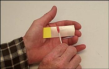
Prepare the smear immediately after withdrawal of the swab by rolling (not sliding or rubbing) the cotton tip along the length of a glass microscope slide. Generally, two parallel tracks can be rolled on a single slide.
As soon as the smear is prepared, dip it 5 to 10 times in a container of methanol, or fix by use of a spray fixative. It is best to fix expeditiously, but allowing the slide to dry fully and remain unfixed for up to a few hours is generally not a problem. After fixation, the slide can be stored for prolonged periods of time, although typically, it is stained without delay.
Diff-Quik is a very convenient stain for use with vaginal smears. It consists of two solutions: an eosinophilic (red) solution and a basophilic (blue) stain. It is best to have a small container of each component and replace the stains with some regularity, as they will become contaminated with vaginal cells after staining a number of slides. To stain the smear, dip the slide in and out of the red stain 5 to 10 times, the in and out of the blue stain 5 to 10 times.
There is no need to rinse the slide between red and blue, but do not dip first into the blue, then into the red, or the red will rapidly become blue! If the staining appears too light or too dark, adjust the number of dips accordingly. A number of other stains can be substituted for Diff-Quik.
After dipping in the blue stain, rinse the slide in tap water and it’s ready to examine. Examine while still wet or dry the slide and apply a coverslip.
Classification of Vaginal Epithelial Cells
Type of cells that we can see in a vaginal cytology
We can classify the cells in keratinized versus nonkeratinized or cornified versus noncornified. The cornification or keratinization is a reflex of circulating estrogens. The basal cells (near blood supply) are small, round and with a clear nucleus, but in the cornified cells (with no blood supply) the nucleus become smaller, pyknotic and finally is disintegrated leaving an anuclear cell.
We will describe the principal characteristics of different types of cells:
Parabasal cells: small, round, slightly oval, large vesiculated nucleus and small cytoplasm. Good staining.
Intermediate cells: slightly larger than parabasal cells to twice that size. Smooth, oval to rounded irregular borders, nucleus smaller than in parabasal cells. More cytoplasm than parabasal cells.
Superficial cells: Are dead cells, are the largest in vaginal cytology with a sharp, flat, angular cytoplasmic borders and a small pyknotic, fading nuclei or without nuclei.
Anuclear squames: irregular vaginal cells, no nucleus, smaller than superficial. These are the cells that have also been called “fully cornified.”
Metestrus cells: Large intermediate vaginal cells that appear to have one or more neutrophils in their cytoplasm. Are typical of bitch diestrus.
Foam cells: parabasal and intermediate cells with obvious cytoplasmic vacuoles. May be associated with diestrus and anestrus.
Neutrophils: Inflammatory cells that can be normal or abnormal in vaginal cytology in function of the estrus period
Red blood cells: Blood can be normal in the bitch but always is abnormal in the queen.
Correlation between ovarian cycle and vaginal cytology
In the bitch
Dogs has two ovarian cycles twice yearly, in spring and again in autumn, however some bitches experience more or less than two cycles a year. These are in function of breed, climate, and others.
The bitch ovarian cycle is divided in four phases: proestrus, estrus, diestrus and anestrus. We can correlate this phases with vaginal cytology.
Proestrus: Period of high follicular activity. The female attracts the males but is not receptive to the copula, the vagina has a bloody discharge and is edematous. The duration of this phase is usually from 6 to 11 days, but the average can be from 2-3 days to 25 days. The vaginal cytology is typical of estrogens secreted by developing ovarian follicles with RBC, numerous parabasal, small and large intermediate vaginal cells, neutrophils are common, bacteria, and a dirty background in the early proestrus and with no neutrophils, less blood cells, more than 80% of superficial cells and a clear background in the late proestrus. This late proestrus is difficult to distinguish from early estrus.
Estrus: In this phase the bitch allows the mating. Estrus starts with this allowing and ends when she no longer accepts the male. Another criterion to define the finish of estrus is the vaginal cytology start of diestrus. Estrogen concentrations reach a peak 1 or 2 days prior to the onset of estrus, and during estrus starts the decline in circulating estrogens and the start in progesterone secretion by luteinized follicular cells first and corpus luteum after. The vaginal cytology is relatively constant with superficial cells and anuclear squames accounting more than 80% of the total vaginal cells and often reaching 100%. No neutrophils must be present and red blood cells are not usually presents.
Diestrus: Is the phase with predominance of progesterone secretion with or without pregnancy. Vaginal cytology is characteristic in this phase with a dramatic “shift” form a estrus with a 80-100% superficial cells to one of 80-100% of parabasal and intermediate cells plus neutrophils and metestrus cells. This change is produced in about 24-48 hours.
Anestrus: Is defined as the period of sexual arrest. The uterus involution is produced and start with whelping and ends with a new proestrus. In non pregnant bitches the beginning of anestrus is not readily easy to distinguish clinically with no obvious demarcation between diestrus and anestrus.
In the queen
Queen reach the sexual maturity after she attains at least 80% of adult body weight and if the photoperiod is appropriate (usually between 6 and 9 months of age). By definition the cat is seasonally polyestrous. She cycles repeatedly throughout a breeding season unless the cycle is interrupted by pregnancy, ovulation, illness or pseudogestation. This is a important difference with the bitch, the influence of photoperiod and hours of daylight.
The phases of the queen sexual cycle are the same that in the bitch, but with some differences. Usually we aggregate proestrus and estrus, because they are difficulty distinguished. Another characteristic of the queen is that she has a interestrual period, between estrus, in the adequate photoperiod season with a typical changes in the vaginal cytology. A typical cycle of a queen is: anestrus-follicular phase (proestrus + estrus)-interestrus-follicular phase ……. – anestrus
Another important characteristic in the physiology of the queen, is that is a ovulation induced female. When the tom introduce the penis in the queen vagina, he induce the ovulation reflex. In some occasions a female can ovulate without copula.
Proestrus: Is the period of follicular function after anestrus or interestrus period, estrogen synthesis, secretion and changes in behaviour and vaginal cytology. The behavioral changes are rubbing, vocalizing, lordosis, and rolling.
Estrus: Is associated with follicular estrogen synthesis and secretion. Usually is associated with proestrus as a “follicular phase.”
Vaginal cytology in “follicular phase”: clear background, without cellular debris and easy visualization of vaginal cells. The squames increase to 10% of the total on the firsts days reaching 40% at seven days, and then slowing again to 10%. In this phase the intermediate cells decrease from 40-50% in the interestrus period to 10% during follicular phase. No parabasal cells, no RBC, and no PMNN are typical of this phase.
Interestrus: follows cessation of follicular activity and the suppression of estrogens secretion. In this phase the behaviour is normal for the queen without rubbing or vocalization. The vaginal cytology is really curious and has a lot of typical characteristics: we can observe all types of vaginal cytology cells, intermediate, squames, parabasal and superficial and again we can see debris in the background.
Diestrus: As in the bitch is the phase with progesterone dominance. The corpus luteum produce progesterone after ovulation (induced ovulation in the queen).
Anestrus: Again, anestrus is defined as a period of clinical reproductive quiescence. The female do not attract males and not display sexual behaviour. In the vaginal cytology we have low cellularity with all possible types of cells and a dirty background with debris.
Clinical usefulness of vaginal cytology
We will see with practical cases the normal and possible applications of vaginal cytology as:
Management of normal breeding
Shipping or receiving a bitch
Study of unusual cycles
Predicting whelping dates
Infertility problems
Diagnosis of vaginal problems, follicular problems and uterine diseases
Application of reproductive drugs: abortifacients
Limitations of vaginal cytology
Vaginal cytology doesn’t permit gestation diagnosis, doesn’t permit identify ovulation day, and is just a complement of a good clinical examination, anamnesis and hormone assays.
A majority of cells observed in a normal vaginal smear are, not surprisingly, vaginal epithelial cells. In addition, varying numbers of leukocytes, erythrocytes and bacteria are usually evident, and small numbers of other contaminating cells and microorganisms are sometimes observed.
Analyzing a vaginal smear is largely an exercise in classifying the epithelial cells into one of three fundamental types: parabasal, intermediate or superficial cells. Keep in mind, however, that the epithelial cells reflect a developmental continuum; some of the cells you observe will not fit perfectly into these rigidly-defined categories.
All of the figures on this page are presented at the same magnification and are from a single dog.
Parabasal Cells
Parabasal cells are the smallest epithelial cells seen on a typical vaginal smear. They are round or nearly round and have a high nuclear to cytoplasmic ratio.
Parabasal cells are prevalent on smears taken during diestrus and anestrus, and not uncommon during early proestrus. Parabasal cells are conspicuously absent during estrus.
Intermediate Cells
Intermediate cells vary in size and shape, but typically have a diameter two to three times that of parabasal cells. Many cytologists subclassify these cells into:

- small intermediates: nearly round or oval shape with large, prominent nuclei
- large intermediates: polygonal shape with a small nuclear/cytoplasmic ratio
In the figures below, all of the cells are typical intermediates except for the one cell in the middle panel (arrow), which might be classified as a superficial cell (small, dark nucleus). Note that the intermediate cells vary in size and that some have rounded outlines (small intermediates), while others have a polygonal shape (large intermediates).
Intermediate cells are prevalent during all stages of the cycle except estrus.
Superficial Cells
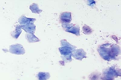
Superficial cells are the largest cells seen on a vaginal smear. The are polygonal in shape and distinctly flat, sometimes having the appearance of being rolled up. Their nuclei are either absent or pyknotic (very small and dark). Superficial cells without nuclei are often referred to as being “fully cornified”.
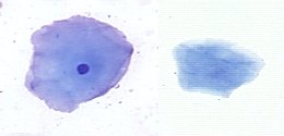
Superfical cells are often seen in large sheets or strings, as seen below with fully cornified cells.

Superficial cells are not normally seen during anestrus and increase in prevalence during proestrus. The presence of large numbers of superficial cells or only superficial cells is the defining characteristic of cytologic estrus, and their abrupt and precipitous decline marks the onset of diestrus.
Other Cells
Aside from the epithelial cells described above, a number of other cells are seen on vaginal smears.
- Erythrocytes are usually observed in large numbers during proestrus. In some bitches, they are seen through estrus and even into early diestrus.
- Neutrophils are often abundant in smears taken during early diestrus, and are not uncommon at other stages, though rare during estrus. Moderate numbers of neutrophils are a common, though not consistent feature of normal canine vaginal smears and not by themselves indicative of vaginitis.
- “Foam cells” is a term given to non-descript epithelial cells containing numerous vacuoles that are typically seen on smears prepared during anestrus.
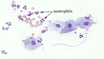
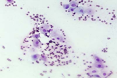
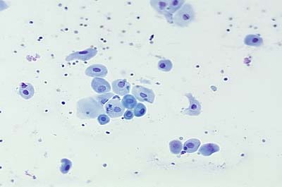
The following figure, of a proestrus smear, shows a group of intermediate cells associated with neutrophils and red blood cells.
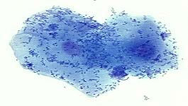
Finally, bacteria are often seen on vaginal smears in huge numbers, covering cells and spilling onto the background. The minute dark specks covering the superfical cells in the image below are bacteria.
Cytologic Changes Through the Canine Estrous Cycle
Stages of the canine estrous cycle can be defined by sexual behavior, physical signs (vulvar swelling, vaginal bleeding) or by vaginal cytology. The period of receptivity to a male varies considerably among bitches; some bitches are receptive well before and after the period of potential fertility. Similarly, signs such as “proestrus bleeding” are often unreliable indicators; some bitches bleed very little and other show bleeding through estrus and into diestrus. Since cytologic changes reflect the underlying endocrine events of the cycle, they are almost always a better predictor of the “fertile time” and gestation length than are behavioral or physical signs.
Cytologic changes through the canine estrous cycle reflect changes in blood concentrations of estrogen. As depicted below, and described in more detail in the section on Canine Reproduction, estrogen levels rise prior to and during proestrus and fall in conjunction with the preovulatory surge of luteinizing hormone. Rising levels of estrogen induce the “cornification” that is characteristic of smears examined during estrus. Ovulation occurs two days after the LH surge.
The sections below describe the cytologic picture typical of different stages of the canine estrous cycle.

Examination of a single smear can sometimes provide useful information, but can also be quite misleading. For example, it is often difficult to differentiate proestrus and diestrus from an isolated smear. It is therefore highly recommended that multiple smears be evaluated.
Anestrus
Intermediate and parabasal cells predominate in smears taken during anestrus.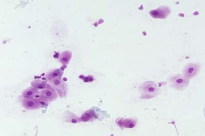
Superficial cells are absent or found in very small numbers. Neutrophils may also be present or absent.
Proestrus
Serum concentrations of estrogen rise during proestrus, leading to capillary breakage and leakage of red blood cells through uterine epithelium, as well as proliferation of the vaginal epithelium. Examination of vaginal smears from early to late proestrus will reveal a gradual shift from intermediate and parabasal cells to superficial cells. Typically, red blood cells are present in large numbers and neutrophils are commonly observed. Large numbers of bacteria are also often present.
In some bitches, proestrus can persist for two to three weeks. In such cases, prolonged lack of receptivity may suggest the need to artificially inseminate or force-breed the animal. Examining vaginal smears in such cases will alleviate such concerns – certainly, if more than a very small percentage of cells are parabasals and small intermediates, breeding is a waste of time.
Estrus
The defining characteristic of cytologic estrus is the predominance of superficial cells. Most, but not all, bitches will undergo full cornification, and the smear will reveal a monotonous pattern composed almost exclusively of anucleate superficial cells.
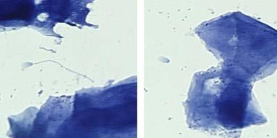
If the bitch has been bred within a day of preparing a vaginal smear, it is quite likely that sperm will be observed among the epithelial cells. Indeed, careful examination for sperm in a smear taken within a few hours of an alleged breeding is a fairly reliable means of confirming or denying such an incident. In the image below, an intact sperm (left panel) and a sperm head (right panel) are present next to superficial cells.
Diestrus
The onset of diestrus is marked by a precipitous decline in the number of superficial cells and reappearance of intermediate and parabasal cells. Most commonly, the cellular profile changes within a single day from essentially 100% superficial cells to less than 20% superficial cells. However, it is best to confirm the onset of diestrus by examining a smear prepared on diestrus day 2.
The significance of identifying the onset of diestrus is that it is a considerably more accurate predictor of the time of ovulation, and hence gestation length, than sexual behavior. Dogs ovulate 5-7 days prior to the onset of diestrus (7-9 days after the preovulatory LH surge), and hence, gestation length is usually 57 + 1 day from the onset of diestrus day 1. The period of behavioral estrus is variable, and often extends up to several days before and/or after cytologic estrus. Gestation lengths calculated from the onset or cessation of receptivity are correspondingly inaccurate. The onset of diestrus also correlates well with loss of fertility, and breedings after the diestrus shift are rarely fertile.
DR. AMIT, CANINE SPECIALIST,PUNE
References and Reviews on Canine Vaginal Cytology
- Concannon PW and DeGregario GB: Canine vaginal cytology. In Burke T (ed), Small Animal Reproduction and Infertility, Lea & Febiger, pp 96-111, 1986.
- Holst PA and Phemister RD: Onset of diestrus in the Beagle bitch: Definition and significance. Am J Vet Res 35:401, 1974.
- Holst PA and Phemister RD: Temporal sequence of events in the estrus cycle of the bitch. Am J Vet Res 36:705, 1974.