BASICS OF TRICHOLOGY: IT’S IMPORTANCE IN VETERINARY FORENSICS
Dr. Munmun Sarma
Professor, Department of Anatomy & Histology
College of Veterinary Science, Assam Agricultural University
Khanapara, Guwahati – 781 022, Assam
Mobile No. 9435047065 Email: munmun.sarma@aau.ac.in
Indian subcontinent is well known for its mega-diversified population of flora and fauna in 2.4% of the world’s land area with 7-8% of all recorded species, including more than 45,000 species of plants and 91,000 species of animals.
Hair in mammals is composed of the hair follicle, root and hair shaft. Follicles are formed only once in the life time of an individual. Physiologically hair fibres form a protective layer over the surface of epidermis protecting the animals from injury, snake bites etc. Hair has the capability to respond to external stimuli and any movement of the fibre is picked up by the hair follicle which is transmitted as a message to the nervous system. The hair fibre is regarded as an “antenna” of the mammals to receive sensory stimuli. Hair also plays an important part in controlling the body heat by providing insulation against sudden heat loss or gain.
Hair is not only important from physiological point of view, but also from forensic aspect as it can help to solve the most complicated vetero-legal cases. Due to human activities like poaching and deforestation many wild mammals are now endangered. Due to this reason, identification of animal species became important in crime investigations in case of poaching and trading of animal parts.
All hairs, whether utilized for textiles or not, have cuticular scales on their surfaces, and most hair (exempting wool) shows presence of a medulla in its centre, the width of medulla is generally found as half or little more of the total diameter of hair shaft (Kirk, 1953). Again, hair can resist putrefaction so it serves as an evidential value where other evidence are either not sufficient or are found unsuitable for crime investigation due to adverse natural environmental conditions.
Therefore, Trichology, an important branch of Biological science that deals with the study of hair which is of utmost importance in Veterinary forensics. The literal meaning of the Word “Trichology” means a branch of medical and cosmetic study of hair and scalp. But when it comes to application of Trichology in Veterinary Forensics one has to understand what is Veterinary Forensics and role of Veterinarians in Forensics. Veterinary forensics is a field of crime investigation wherein scientific knowledge is applied to collect and examine evidence from crime scenes where animals have been killed, particularly those that have been protected by Law. Whether a Vet has been engaged in domestic or wildlife crime investigation, one has to be very careful during the crime investigation and meticulously analyse the evidence before drawing any conclusion while preparing the report.
Animals have three types of hair (Dyce et al., 1996):
- Guard hair: straight and rather stiff outer hair, provide a top coat to the underlying fur coat of the body,
- Wool hair: fine and wavy, provide a undercoat of the body
- Tactile hair: stout and have restricted distribution and is responsive to mechanical stimulation.
A single hair on a macro-scale has –
(1) A root,
(2) A shaft, and
(3) A tip.
The root of hair is the proximal most portion of the hair and is in the follicle.
The hair shaft is the main portion of the hair that projects above the skin.
The hair tip is the distal most portion of the hair.
Basic components of hair are –
- Keratin (a protein),
- Melanin (a pigment), and
- Trace quantities of metallic elements.
There are three main structural elements in a hair from outside to inside are –
- The cuticle,
- The cortex, and
- The medulla.
Hair cuticle or cuticular scale pattern is mainly of three types—
- coronal (crown-like),
- spinous (petal-like), and
- imbricate (flattened).
The coronal, or crown-like scale pattern, is found in hairs of very fine diameter and resemble a stack of paper cups, for example hair of Tiger (Fig. 1), small rodents, bats and rarely in human hairs.
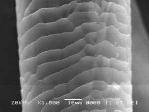
Fig.1: SEM photomicrograph of Coronal cuticular scale pattern of Tiger hair
Spinous or petal-like scales are triangular in shape and protrude from the hair shaft, found in mink hairs and on the fur hairs of seals, cats, and some other animals. They are never found in human hairs.
Imbricate scales – cuticular scales are overlapping with narrow margins, commonly found in human hairs and many animal species eg. Mithun (Fig.2)
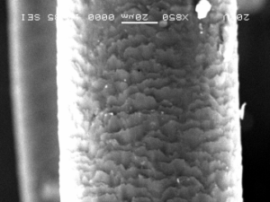
Fig.2: SEM photomicrograph of imbricate cuticular scale pattern of mithun hair
Hair cortex: Hair cortex (Fig.3) is made up the bulk of the hair. It is the mainly composed of elongated and fusiform (spindle-shaped) cells.
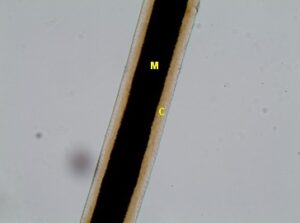
Fig.3: Hair histology showing cortex (C) and medulla (M) of tiger hair
Other structures present in hair are-
- Cortical fusi: Cortical fusi are irregular-shaped airspaces present in hair which may be of varying sizes
- Pigment granules: Pigment granules are small, dark, and solid structures which are granular in appearance commonly found in cortex.
- Large oval-to-round-shaped ovoid bodies: Ovoid bodies are large (larger than pigment granules) are solid structures that vary from spherical to oval in shape, with smooth margins. They are abundantly found in hair of some cattle and dog as well as in other animal hairs.
Hair medulla: Hair medulla is composed of round cells in case of thick or coarse hair (guard hair of animals and usually absent in fine hair or down hair. Presence or absence of medulla is species specific and in some species of animals it is found to be gender specific. In some species medulla is composed of sparse cells and air bubbles. Hair medulla constitutes the central core of hair. When filled with air, it appears as a black or opaque structure under transmitted light and when filled with mounting medium or some other clearing substance, the structure appears clear or translucent in transmitted light, or nearly invisible in reflected light. In human hairs, the medulla is generally amorphous in appearance, whereas in animal hairs, its structure is frequently very regular and well defined.
Gross examination of hair strands of animals show three different types of medulla-
- Fragmented or trace,
- Discontinuous or broken, and
- Continues type.
Micro-morphologically there is different type of hair medulla found in animals. They are-
- Uniserial ladder (Fig.4),
- Multiserial ladder (Fig.5) ,
- Cellular or vacuolated type
- Lattice type (Fig.6)

Fig.4 : Photomicrograph of Uniserial-Ladder Medulla of a rabbit
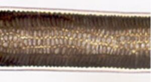
Fig.5 : Photomicrograph of Multiserial-Ladder Medulla of a rabbit

Fig.6 : Photomicrograph of Lattice type medulla found in deer
Other parameters of hairs that are to be studied to solve crime investigation are-
- Length of hair
- Colour of hair
- Diameter of shaft
- Thickness of cortex
- Diameter of medulla
- Medullary index as per Kirk (1953)
- Cortico-medullary index as per Kirk (1953)
Importance of guard hair in crime investigation–
- Being the outermost hair coat of the animals, it is mostly left behind in the scene of crime by the criminals.
- Guard hair make good forensic evidence because they can survive for many years, they carry lot of biological information, and are easy and effective to examine.
- Hair is chemically most stable, resist putrefaction and retain its morphological characters for a long duration/ period.
- Retains their uniqueness despite of processes like ageing, digestion and change in the environment
Veterinarians Role in Crime investigation (Forensics):
- Veterinary forensic experts convert the clues collected from the scene of crime into evidence admissible in the court of law.
- Any biological samples or skeletal parts provided by Police Department, Forest Department, and Excise Department etc are to be properly investigated before submitting the report to the court for conviction of the criminal.
- To ascertain the cause of death of an animal suspected to be poisoned or diagnosis of diseases, necropsy should be carried out following standard post mortem protocol.
Very often forensic samples like meat and guard hair of animals are sent for species identification and these forensic samples are mostly from fringe areas of Wildlife National Parks, Wildlife National Sanctuaries and wildlife protected areas. Now the matter is how smartly the Veterinary forensic expert can act and prepare the report that is to be submitted in the Court of Law for conviction of the criminals indeed.
Many a times the author had to address such issues case-by-case to solve various vetero-legal cases and one such case is being discussed as to how such cases should be handled by a Veterinarian (location of incidence is not mentioned as the case is in the court). The story behind the incident was that a meat seller was selling pork (pig meat) in the open market which happened to be suspected as meat of wild pig. This information immediately reached the concerned authority, the meat was confiscated by the concerned authority and the seller was kept behind bar. The meat sample with hair strands adhering to the meat was sent for the species identification. Since it was a vetero-legal case and the report had to be sent to the Hon’ble Court of Law for conviction of the suspect, the author had to carry out the different parameters step-by- step, the procedure discussed hereunder for preparation of an authentic report. The first step was macro-morphological study of guard hair stands.
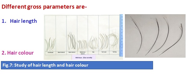
Macro-morphological or gross study of the hair strands gave the clue that it was of a wild pig (Fig 7).
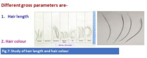
In the above slide (Fig.7), the hair strands on the right slide showed split ends which were a gross characteristic of hair of wild pigs and similar features were shown in the slide on the left which is taken the data base prepared by the author in the departmental laboratory.
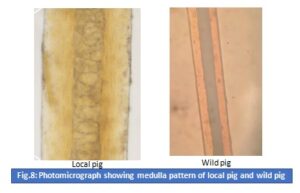
Some hair strands of the forensic sample and few hair strands were domestic/ local pig of Assam were processed for micro-morphological study and their medulla pattern was compared with the database which showed that the medulla pattern showed the characteristics of wild pig (Fig. 9).
As the culprit was kept behind bars therefore, for conviction of the offender, mtDNA sequencing from hair bulbs was done (Figs. 9= 1, 10) to ascertain accuracy in the report that was sent to the Court of Law.
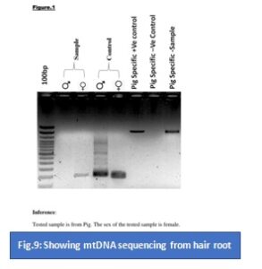
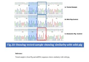
From the above study it was confirmed that the forensic sample was that of wild pig.
With the quote of the famous chemist late Paul L. Kirk, from Crime Investigation let me conclude-
“Wherever he steps, whatever he touches, whatever he leaves, even unconsciously, will serve as a silent witness against him. Not only his fingerprints or his footprints, but his hair, the fibres from his clothes, the glass he breaks, the tool mark he leaves, the paint he scratches, the blood or semen he deposits or collects. All of these and more bear mute witness against him. This is evidence that does not forget. It is not confused by the excitement of the moment. It is not absent because human witnesses are. It is factual evidence. Physical evidence cannot be wrong, it cannot perjure itself, it cannot be wholly absent. Only human failure to find it, study and understand it, can diminish its value.”
REFERENCES
Dyce, K. M., Sack, W. O. and Wensing, C. J. G. (1996): Text Book of Veterinary Anatomy. Second Edition. Published by W. B. Saunders Company. Philadelphia London New York St. Louis Sydney Toronto
Kirk Paul K. (1953): Crime Investigation Physical evidence and the Police Laboratory. Published by John Willey & Sons. New York London Sydney Totonto
https://www.pashudhanpraharee.com/category/toxicity/
https://www.ncbi.nlm.nih.gov/pmc/articles/PMC6088218/
*****************************************************************************


