Canine Transmissible Venereal Tumour (CTVT) or Transmissible Venereal Tumour (TVT):Treatment & Management
Dr-Amit Bhardwaj ,Veterinary Surgeon , Goa/Pune
Canine transmissible venereal tumour (CTVT), also known as transmissible venereal tumour (TVT) or Sticker’s sarcoma, is a transmissible cancer that affects dogs. CTVT is spread by the transfer of living cancer cells between dogs, usually during mating. CTVT causes tumours which are usually associated with the external genitalia of both male and female dogs.It also affects other canids such as coyotes, foxes and jackals.
Also known as infectious sarcoma, venereal granuloma, transmissible lymphosarcoma or Sticker tumor, this disease is a benign tumor that occurs primarily on the external genitals of both male and female dogs. It is one of a very few tumors that can be transmitted by direct contact. It acts like a freely living organism — more a parasite than a cancer.
Canine transmissible venereal tumour (TVT) or Sticker’s sarcoma was first described by Hujard in 1820 in Europe and its name has been associated with Sticker who systematically studied it in the beginning of the 20th century. It is a neoplasm with unusual properties and unconventional clinical development, naturally occurring exclusively in dogs contaminated primarily by sexual contact and possibly by direct contact related with social behaviour (e.g., sniffing, licking of the genitalia, bite wounds during fights). It is almost always located at the genitalia of both sexes and is rarely found elsewhere in the body, while it metastasises in only a very few cases. Transmission occurs by inoculation of intact neoplastic cells in the damaged mucosa or skin.
https://www.pashudhanpraharee.com/treatment-management-of-transmissible-venereal-tumortvt-in-dogs/
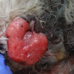
Canine TVT is cauliflower-like, pedunculated, nodular, papillary, or multilobulated in appearance. It ranges in size from a small nodule (5 mm) to a large mass1. Finding a small nodule that bleeds and is located on the external genitalia is the most consistent symptom. The condition is transmitted during sexual contact and is most commonly seen in young, but mature, sexually active animals.
AETIOLOGY AND TRANSMISSION
The transmission of TVT by means of intact neoplasm cells was assumed already from the beginning of the 20th century (Sticker 1905). Later, it was proven that the dog is the only host of TVT (Bloom 1954). Then it was confirmed that the mutated neoplasm cell itself was the causal agent of TVT and that it induces an immune response in the host (Cohen 1972, 1978). Significant morphological differences have been repeatedly demonstrated to exist between normal dog cells and cells of the tumour. These include constantly some highly specific chromosome aberrations, which, when associated with other factors, lead to the conclusion that TVT derives from cells that have undergone a mutation caused by a still unknown factor. Tumour cells are exfoliated and transplanted during coitus from animal to animal and perpetuate themselves like any other heterologous, mono-cellular organism. The mechanism allowing the neoplastic cells to override nature’s histocompatibility barrier is unknown, while the presence of active immunity has been demonstrated.
Symptoms of Transmissible Venereal Tumor in Dogs
The main symptom of this disease is the presence of tumors which are usually located in the genitals of both sexes, as well as the nasal and oral cavities. While tumor spread is uncommon, it can occur without a genital tumor being present. Locations can include:
- Mouth
- Anus
- Skin
- Lymph nodes
- Pharynx
- Tonsils
- Eyes
- Liver
- Muscles
- Kidney
- Spleen
- Brain
Tumors can appear as small papules or nodules, and over time, they can progress into a cauliflower-like, multi-nodular, or multi-lobulated appearance. They can range in size from 5 mm to 15 cm in diameter, and are usually firm. Tumors often ulcerate, become inflamed, and bleed easily. Other signs associated with these tumors include:
- Bloody discharge from vaginal areas in females
- Bloody discharge from penis in males
TREATMENT
At present, surgical treatment, chemotherapy and radiotherapy are used to treat TVT, while immunotherapy has not been proven effective.
Surgical treatment has been applied since the last century (Wong & K’Ang 1932) with a low rate of efficacy (30-35% relapses due to tumour cell transplantation into the surgical wound during operation). The use of electrocautery makes the operation easier and seems to be a little more effective; however it is still far from being suggested as the first choice. Therefore, surgical treatment might be applied to those dogs that present solitary, small, easily accessible and noninvasive tumour nodules.
TVT has been proven to be highly sensitive to irradiation, already from the last century, (Wong & K’Ang 1932). Solacroup proposed radiation therapy for TVT in Europe in 1950, which has been employed since, mainly in France resulting in a sufficient up to date incidence reduction of TVT cases. Dosage recommendations range from 1500 to 2500 rads (depending on the chronicity and the extension of neoplastic lesions), divided in sessions of 400-500 rads over a period of 1-2 weeks, or a single dose of 1000 rads which, if not curative, can safely be repeated 1-4 times. However, radiotherapy lacks practicality due to requirements like trained personnel, specialised equipment and expenses. Therefore its use is recommended in cases where other treatments fail.
Sticker was the first who attempted TVT chemotherapy (arsenicals) at the beginning of the 20th century. The trials with new generation–antineoplastic drugs started during the 6th decade (Deschanel 1962) and vincristine, which was destined to become the drug of choice for TVT therapy, was included in treatment protocols since the 8th decade (Broadhurst 1974).
Currently, the intravenous administration of vincristine at the dose of 0.6 mg/m2 of body surface, once a week, for 2-6 weeks, is the treatment of choice, irrespective of a) the neoplasm size–extent; b) the presence of metastases; and c) the duration of the disease. The time needed to complete treatment and the expenses involved are within reasonable limits. The animals fully recover, with no impact on behaviour and reproductive ability.
Based on our experience, we can raise the following interesting points:
Dogs being infected for less than 1 year, i.e., TVT at initial stages of progression are easily treated. The presence of metastasis, the gender or the age of the animals treated do not influence the duration of chemotherapy. Metastasis subsidence concurs impressively.
Chronic cases infected for more than 1 year may resist to treatment, thus demanding therapy of longer duration without ensuring successful results. In case of failure, radiotherapy gives excellent results; alternatively doxorubicin chemotherapy may be applied.
The potential for a successful vincristine treatment becomes markedly limited when treatment course is interrupted for 2 weeks or more.
The presence of small size tissue remnants (of less than 0.5 cm), which do not bleed after being rubbed, must not be a reason to continue treatment.
Temporary side effects (partial anorexia, mild depression) may be reported in less then 20% of the treated dogs, usually 1-2 days after vincristine administration. Chemotherapy may cause a decrease of the white blood cell count (transient leukopenia), but only a few cases (less than 2%) present such a leukopenia that might deserve an adjunctive antibiotic treatment or impose suspension (discontinuation) of one or more chemotherapeutic administrations.
Before initiating vincristine chemotherapy, it is important to assess the general health condition of the animal while during therapy it is necessary to follow up the total number of leukocytes, at least at weekly intervals.
Reference-On Request


