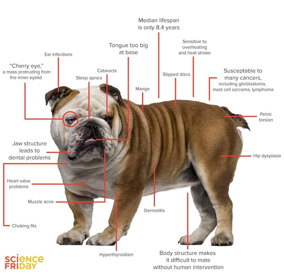
DEGENERATIVE VALVE DISEASE IN ENGLISH BULLDOG
Compiled & Edited by-DR RAJESH KUMAR SINGH, JAMSHEDPUR, 9431309542
Degenerative valve disease is a condition that can affect certain breeds of dogs, including English bulldogs. Staying informed of the symptoms, causes, treatment and prognosis will help you determine what to do in the unfortunate event that your English bulldog develops this condition
Degenerative valve disease accounts for 75 percent of cardiovascular disease in all dogs. Numerous dog species are affected by degenerative valve disease, though the disorder primarily affects dogs classified as small and medium in size, including English bulldogs. While the disease is very well understood, treatment options are limited.
Degenerative Valve Disease
Degenerative valve disease is a heart condition that develops as a result of thickening within the mitral valve of the heart. The thickening causes accumulation of blood within the heart, which leads to increased pressure in the heart. It is breed- and age-specific. The English bulldog breed is at risk of several heart conditions, including mitral valve defects and other conditions that prevent the heart valves from opening properly.
PHYSIOLOGY & CAUSES OF DEGENERATIVE VALVE DISEASE
Although the actual cause of degenerative valve disease is not known, it is characterized by nodular thickening of the cardiac valve leaflets. This thickening causes blood to accumulate in the heart, causing increased pressure
Chronic degenerative heart valve disease (CVD), technically known as endocardiosis, is the most common clinically significant heart disease of dogs. While any dog may be affected, it is most prevalent in older small breed dogs. The disease may begin to develop within the first few years of life, but usually does not become clinically apparent until the later years; there is substantial variation in this regard between breeds and individuals. The cause of the condition is not known but there are clearly genetic factors that predispose to early-onset and/or more severe disease. As such this is an “acquired” heart disease that cannot be detected in the early years of life, and yet genetic factors are present throughout life that predispose to the condition.
CVD can affect any of the 4 heart valves, but clinical disease is most apparent affecting the mitral valve by a wide margin. The mitral valve is a marvelous structure that admits oxygenated blood returning from the lungs into the main pumping chamber of the heart, the left ventricle, and closes when the muscle of the ventricle begins to contract. The left ventricle generates the pressure and blood flow through the aorta needed to supply the entire body with nutrients and oxygen. Normally the leaflets of the mitral valve are smooth, and flexible; the normal valve is so thin that you would be able to read a printed page through it and so efficient that there is no detectable leakage whatsoever. CVD affecting the mitral valve causes the valve leaflets to become thickened and irregular so that the valve leaks. Subsequently some of the blood is ejected backwards through the leaking valve into the left atrial chamber each time the left ventricle contracts. Therefore the severity of the CVD condition is a largely dependent on the degree of leakage. The leaking mitral valve requires the left ventricle to pump blood in TWO directions, “forward” into the aorta and “backward” into the left atrium.
This is a condition that develops typically over years of a dog’s life. Most commonly the first indication of a problem is from the detection of a heart murmur by the veterinarian. A heart murmur in this case is caused by abnormal blood flow through the leaking mitral valve which makes a sound that is detectable with a stethoscope. The murmur is caused by the turbulence of the blood flow through the abnormal valve, similar to water flowing over rapids as opposed to smooth flow in a river. For the mitral valve there is a relatively strong association between the loudness of the heart murmur and the severity of the disease since the murmur becomes louder with greater leakage of the valve. The loudest heart murmurs produce a vibration on the chest wall called a thrill that can be determined by touch (“palpation”) without the aid of a stethoscope. Another indication of severity that may be apparent on a physical examination is a pounding of the heart against the chest wall. As the disease worsens, the left ventricle must literally pump more since it ejects blood both into the aorta plus an additional amount through the leaking mitral valve into the left atrium. With increasing disease severity the heart literally changes its structure, enlarging to accommodate the need to pump more blood.
The onset of a heart murmur in an older dog warrants evaluation of the patient by physical examination, blood pressure determination, and either a chest x-ray at the generalist’s office or an echocardiogram performed by a cardiology specialist. Chest x-rays provide good information about the size of the heart and are best for evaluating the lungs if there are any respiratory symptoms (e.g. cough, difficulty breathing). An echocardiogram is an ultrasound study of the heart and provides superior information about the size and function of the heart, the cause of any heart murmur, and the severity of mitral valve leakage in particular. An echocardiogram also provides information relating to complications of the condition such as elevated blood pressure in the lungs (pulmonary hypertension), and whether more than one valve may be affected, an aspect that may be obscured when there is a loud murmur from the mitral valve.
The left atrium is the chamber that holds the oxygenated blood returning from the lungs and has an important “reservoir” function. With increasing severity of mitral regurgitation, the jet of blood swirls with increasing intensity in this chamber and the pressure builds. Consequently the left atrium is something of a “barometer” for assessing the disease severity.
Signs and Symptoms
In the early stages, English bulldogs may not experience visible symptoms. In later stages, they can suffer from weight loss, fainting, increasingly difficult breathing or increased coughing. Unfortunately, because heavy or labored breathing is common in English bulldogs, it can be challenging to distinguish normal breathing from that associated with degenerative valve disease. Because of this, along with the fact that mitral valve disease is common in bulldogs, it’s best to visit your vet for regular checkups. Several symptoms are indicative of degenerative valve disease, including labored or heavy breathing, increased coughing, fainting spells, restlessness and weight loss. Symptoms like labored breathing, which is common in bulldogs, are sometimes difficult to diagnose.
Diagnosis
A vet can detect a heart murmur even in early stages without your bulldog experiencing symptoms. To help detect a problem, the vet will listen for respiratory symptoms like wheezing and lung crackling. In addition, she might conduct an echocardiogram, a type of ultrasound, of the heart. This can help reveal thickening, enlargement or irregularity in the shape of the heart’s valves. This noninvasive procedure entails attaching sticky patches to your pup’s body and reading the electrical currents within his heart.
Prevention, Treatment and Prognosis
At this time there are no proven prevention methods for degenerative valve disease. There are several ongoing clinical trials, though none have proven effective at slowing or preventing the disease., no prevention protocol for degenerative valve disease for English bulldogs exists. Treatment generally focuses on combating congestive heart failure, which is often the long-term end result of the disease and the heart conditions that potentially plague the bulldog breed. A vet might prescribe drugs including diuretics, enzyme inhibitors or others, along with periodic removal of fluid within your English bulldog’s heart. Survival rates vary, and can average about a year with therapy. Long term, degenerative valve disease typically results in congestive heart failure in English bulldogs
.
Treatment
As noted above, the cause of CVD is not known and unfortunately there is no treatment known that will either halt or reverse the degeneration of the valve. In humans with sufficiently severe mitral valve disease, open-heart surgery is often performed to repair or replace the damaged valve. Unfortunately this approach is largely unfeasible for dogs at this time due both to the expense of the procedure and the much smaller size of the patients. Advances are being made that may soon allow corrective treatment using interventional methods by which a functional valve may be inserted into position through a blood vessel, without having to perform a major surgery. These methods are not yet widely available for dogs however and current treatment is largely medical and directed at the symptoms and problems caused by the disease.
Veterinary cardiologists have recently published guidelines for the diagnosis and treatment of worsening CVD. Dogs in the early stages of CVD generally feel well and exhibit no symptoms. It’s uncommon for pet owners to be aware of the condition until informed by their veterinarian that their dog has a heart murmur. The heart is actually normal in size and anesthetic procedures such as dental prophylaxis can be undertaken with safety. There is often no need to change any aspect of the dog’s care although we do recommend a balanced diet and avoidance of excessive sodium at this stage. Lifestyle changes that affect overall health may include weight reduction in conjunction with regular exercise, something we can all benefit from. Elevated blood pressure may be expected to worsen leakage of the mitral valve so attendance to any circumstances that promote hypertension (elevated blood pressure) is advocated.
With increasing severity of the disease, the mitral valve leak worsens and the heart begins to enlarge. This stage encompasses a wide range of disease severity as the heart gradually enlarges to accomodate the need to pump blood both forward and also backwards through the leaky valve. Owners may notice their pet “slowing down”; there may be decreased interest in the long walks or dogs may balk at the idea of moderate to intense exercise. When sufficiently enlarged, the heart actually begins to press on the bronchial tubes of the lungs and a cough may ensue. Canine patients may begin to exhibit signs suggestive of a breathing problem including a gagging cough that’s often most apparent after a period of inactivity or sleep. The heart disease may be accompanied by other old age conditions affecting the respiratory tract, sometimes making it difficult to determine the cause of respiratory symptoms. While progression usually occurs over years, it is also possible for sudden worsening of symptoms to occur as a result of a sudden change in the valve structure and function. Veterinary cardiology specialists don’t all agree on exactly what treatments should be initiated, and when. Suffice it to say that treatments are primarily symptomatic and taylored to the individual’s needs and comfort. Anesthesia increases in risk throughout this stage of the disease.
With further progression of the disease, pressure increases in the left atrium and in the blood vessels of the lungs to an extent that may literally push the liquid portion of the blood through the wall of the blood vessels. Accumulation of this fluid within the tissue or air spaces of the lung is known as congestive heart failure (CHF) and is a sign that the condition is quite advanced. At this stage of the disease there are clearly treatments that improve both quality and length of life. However heart failure whose cause cannot be reversed is regarded as a terminal condition; this is true of humans as well as dogs. Average life expectancy is something less than a year after the onset of CHF although there is wide variation among individuals. Standard medical treatment in this phase includes a drug that increases the strength of heart muscle contraction called pimobendan (trade name Vetmedin). Diruretic drugs such as furosemide (trade name lasix) help the patient to rid the body of excess salt and fluid; the latter is a hallmark of severe heart disease and is one of the reasons for fluid accumulating in the respiratory tract. Diuretics increase urination, thirst, and may potentially promoted loss of electrolytes from the body that may require counteractive treatment (sodium, potassium, and chloride). A third standard treatment consists of a class of drugs called angiotensin converting enzyme inhibitors. ACE inhibitors block the production of angiotensin which is chemical produced in the body that’s involved in blood pressure control, causing constriction of blood vessels and further promoting retention of salt and fluid. In the late stages of disease, avoidance of excess sodium in the diet is also quite important and there may be other treatments essential for individual comfort.
Dosages of these powerful medications are carefully adjusted as the disease worsens to optimize patient comfort and well-being. With proper medication, dogs appear comfortable and happy in most cases, even when disease is severe. However there comes a time when drug dosages cannot be increased further, to lessen heart failure symptoms, without causing significant problems from the medications themselves.
NB-This information is not meant to be a substitute for veterinary care. Always follow the instructions provided by your veterinarian.
Reference-On Request


