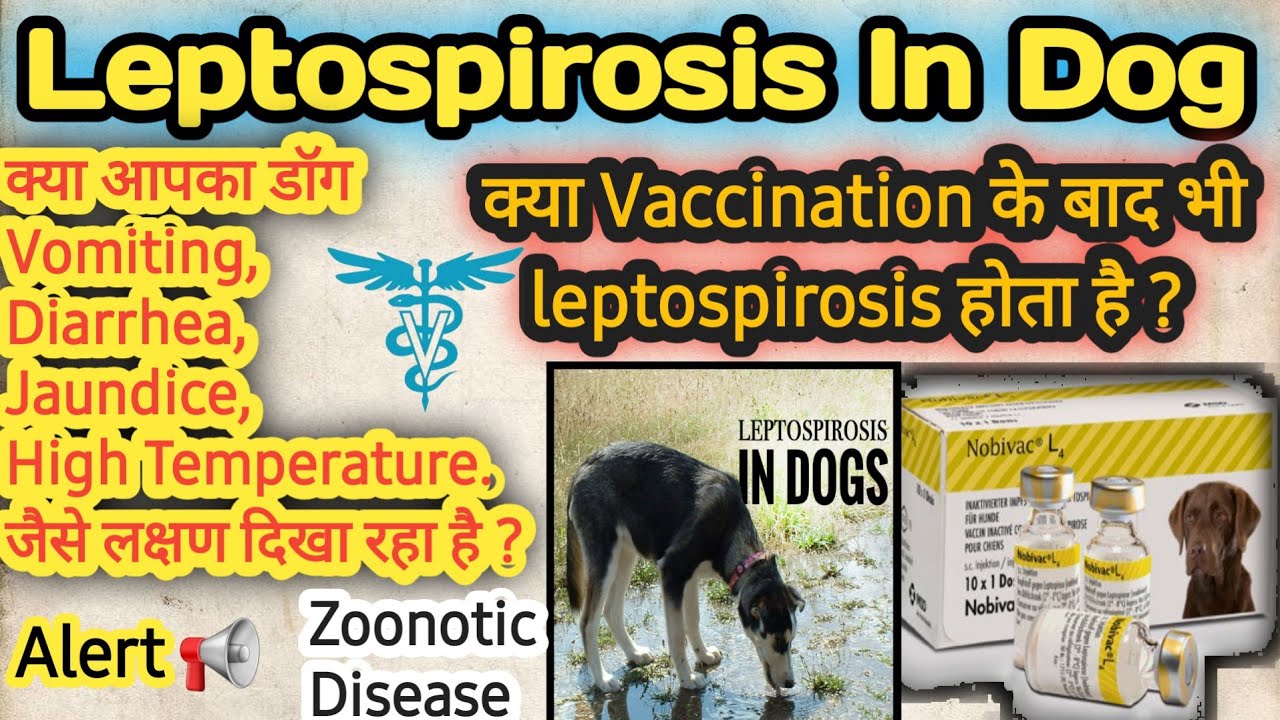
Diagnosis and Treatment of Leptospirosis in a Dog
DR AMIT ,CANINE SPECIALIST,PUNE
Leptospirosis continues to be a significant clinical presence in canine medicine. In addition to an increased number of cases, more diverse clinical presentations are being recognized. Selection of appropriate vaccines and inter-pretation of serological results in the presence of vaccinal titers are emerging issues in clinical practice. Leptospirosis is caused by Leptospira are thin, filamentous, aerobic spirochete bacteria measuring approximately 6-12 µm long. More than 200 serovars of leptospira were identified worldwide; most of them are pathogenic in dogs including Leptospira serovars bratislava, canicola, icterohemorrhagica, pomona, and grippotyphosa. Infected animals become bacteremic and leptospira organisms multiply in the kidney, liver, spleen, central nervous system, ocular tissue and genital tract. In dogs, serovars canicola and grippotyphosa results in more renal dysfunction, whereas serovars icterohemorrhagiae and pomona produce more hepatic damage .Reservoir hosts may be sub clinically infected and shed organisms for months to years after recovery.Leptospirosis, commonly called lepto, is a bacterial infection that can affect dogs, humans, and other mammals. Many strains of Leptospira bacteria can infect dogs.Lepto can cause liver and kidney damage, and depending on the severity of the illness, permanent dysfunction of those organs could occur.Dogs that become infected with Leptospira bacteria may not show any signs of illness. Sometimes, the infection can be cleared by the body without causing disease. However, dogs that do become ill may show some of the following signs:
- Fever
- Trembling
- Lameness caused by muscle or joint pain
- Hiding or difficulty or reluctance to move
- Decreased or absent appetite
- Increased thirst and urination
- Vomiting
- Diarrhea
- Jaundice (yellow skin and mucous membranes)
- Swollen legs
Etiology
- Infection with pathogenic Leptospira Leptospira spp serovars. Several different serovars reported in canine disease.
- Leptospira spp have worldwide distribution.
- Leptospira spp are inactivated by acidic urine, direct sunlight, temperatures above 30°C, desiccation and disinfection. They survive in contaminated water, stagnant or slow-moving water, moist soil; they prefer alkaline pH. Freezing can decrease the survival. Consequently, the disease occurs more common in summer and autumn/fall.
- Each serovar is believed to have a reservoir or maintenance host in which it can cause a chronic infection without overt clinical signs.
- Leptospira spp commonly sequestered in the renal tubules and voided in the urine.
Predisposing factors
General
- Large breed, male adult, outdoor dogs are most commonly infected.
- Infection occurs commonly through urine-contaminated water.
- Likelihood of infection increases in persistent rainfall and areas of poor drainage or contact with slow-flowing water.
- High density of reservoir animals (rodents), or other wildlife are risk factors.
- Herding dogs, hunting dogs, and working dogs are predisposed.
- Skin trauma allows organism to penetrate easier.
Specific
- Unvaccinated dogs have a higher risk of infection.
Pathophysiology
- Infection occurs through ingestion of infected rodents or penetration of mucosae or traumatized skin. Leptospiremia occurs within 1 week. Leptospires spread to other organ systems (kidneys, liver, spleen, endothelial cells, lungs, uvea/retina, skeletal and heart muscles, pancreas, and genital tract) and cause tissue damage, visceral and vascular inflammation.
- Leptospiral pulmonary hemorrhage syndrome (LPHS) can occur as severe manifestation of acute leptospirosis.
- Leptospires can persist in immune privileged site (eg, renal tubes, eye).
- In the presence of adequate antibody titers, leptospires are eliminated from most organs. In the presence of low antibody titers mild leptospiremia can continue with a subclinical course of disease.
Time course
- Time course varies according to immunocompetence of the host, infecting dose, and serovar.
- Incubation period lasts approximately 7 days after experimental infection.
- Initial leptospiremia lasts 4-12 days.
- Usually clinical signs have acute onset.
- Death can occur within 2 days (or later).
- Surviving animals can shed the organism in urine for up to 3 months (or longer).
- Chronic renal sequelae are possible in recovered dogs.
Epidemiology
- Infected reservoir hosts shed leptospires with urine. This leads to infection of further reservoir hosts or infection of accidental hosts, such as dogs and humans.
- Cats can be infected and also shed leptospires and thus, can potentially infect dogs or humans.
Diagnosis of Leptospirosis in Dogs
Your veterinarian will first perform a thorough physical exam and ask you many questions about the course of your dog’s illness. If the doctor suspects leptospirosis infection at that point, some testing will be done to look for coinciding lab results. Those tests might include:
- Urinalysis
- X-rays
- Ultrasound
- Blood tests
Therapy
In canine leptospirosis, renal and liver failures are potentially reversible and should be treated as early and aggressively as possible. The affected dogs are treated symptomatically with antiemetics and gastric protectants, and particular attention is paid to adequate urine production after the animals have been properly rehydrated. The placement of a sterile urinary catheter can be helpful in assessing urine production as well as containing potentially infective waste. Urine production < 2 ml/kg/hr in an adequately hydrated dog indicates oliguria and must be treated aggressively. In animals that are not fluid overloaded, mannitol is usually considered the treatment of choice. It is initially given as a bolus, (0.5 g/kg over 30-60 min) and then followed as a constant rate infusion (1-2 mg/kg/min) if urine production responds appropriately. Alternatively, furosemide can also be administered. It is initially given as a bolus (2-4 mg/kg) and then followed as a constant rate infusion (0.25-1 mg/kg/hour) if urine production increases appropriately. Urine production should be followed closely and over-hydration of the patient avoided. Antibiotic therapy is usually given in 2 phases: ampicillin or amoxicillin can be administered parenterally (20-25 mg/kg i.v. TID) during the initial, critical phase. It is important to note that the kidneys clear these drugs and blood concentrations can become inappropriately high in patients with renal dysfunction. A common method of adjusting these antibiotic dosages is to multiply the normal dose by 1/serum creatinine. When the dogs are recovered, doxycycline (10 mg/kg p.o. daily in 1 or 2 doses) is the antibiotic of choice and is prescribed for a minimum of 3 weeks to prevent persistent renal shedding. The prognosis of canine leptospirosis is fair; depending on the case series, between 50 and 90% of dogs can be discharged from the hospital after a stay of up to 7-10 days. Oliguric/anuric renal failure is a strong negative prognostic factor in most reports. However, dogs affected with this complication do benefit from hemodialysis or continuous renal replacement therapy. Both procedures may significantly decrease mortality, and are currently available in many teaching hospitals and large referral centers. Generally The animals are treated with streptopenicillin @ 40,000 IU/kg body wt. Other supportive therapy may be included imferon 1ml i/m, neohapate 2ml i/m, rantac 1.5ml s/c, and stadren 2ml i/m. Fluids are also given during the five day treatment period.
Prevention
Currently, the mainstay of prophylaxis consists of vaccination of dogs at risk with a bacterin containing the 4 main serovars (canicola, icterohaemorrhagiae, grippotyphosa and pomona). It is not known if this vaccine causes cross-protective immunity against other serovars such as autumnalis, bratislava and australis. In kennels in which leptospirosis is a problem, the environment must be optimized to avoid risk factors.
Zoonotic Potential
Urine from dogs with leptospiruria can infect people if it comes in contact with mucous membranes or skin lesions. In industrial countries, most infections in humans are associated with occupational exposure to infected wildlife or domestic animal hosts, or practice of water sports activities. Although urinary excretion of leptospires ceases shortly after administration of systemic antibiotics, it is recommended to take precautionary measures with hospitalized infected dogs to avoid contamination of the staff. Latex gloves (and long sleeve shirts) should be worn when handling these dogs in the initial phase of treatment. Disinfection of contaminated areas should be done with goggles and face masks. It is important to remember that healthy dogs excreting leptospires in the urine represent a higher risk for human infection.
REFERENCE-ON REQUEST
https://www.pashudhanpraharee.com/leptospirosis-in-dogs/


