


DOURINE: A VENEREAL DISEASE OF EQUINE
Aditya Kumar1, Simran jeet Singh2*, Abha Pant3 and Priyanshi Dobal4
¹Ph.D Scholar, Department of Veterinary Medicine
2PG Scholar, Department of Veterinary Medicine
3,4UG Scholar
College of Veterinary and Animal Sciences
G.B. Pant University of Agriculture and Technology, Pantnagar, Uttarakhand, India- 263145
Corresponding author mail- simranjeets140@gmail.com
INTRODUCTION
Trypanosoma equiperdum is classified under subgenus trypanozoon which also includes other species like T.evansi, T.brucei spp. However, this classification remains controversial as there are limited number of T. equiperdum reference strains. T.equiperdum is the only trypanosome transmitted during sexual intercourse and causes Dourine also called as Dollar’s disease in equines. It is a contagious disease of breeding solipeds characterized by swelling in the external genitalia as well as accompanied by systemic signs include fever, lethargy, anorexia, along with skin lesions. T.equiperdum cannot survive for long outside the body of the host and is not transmitted by fomites.
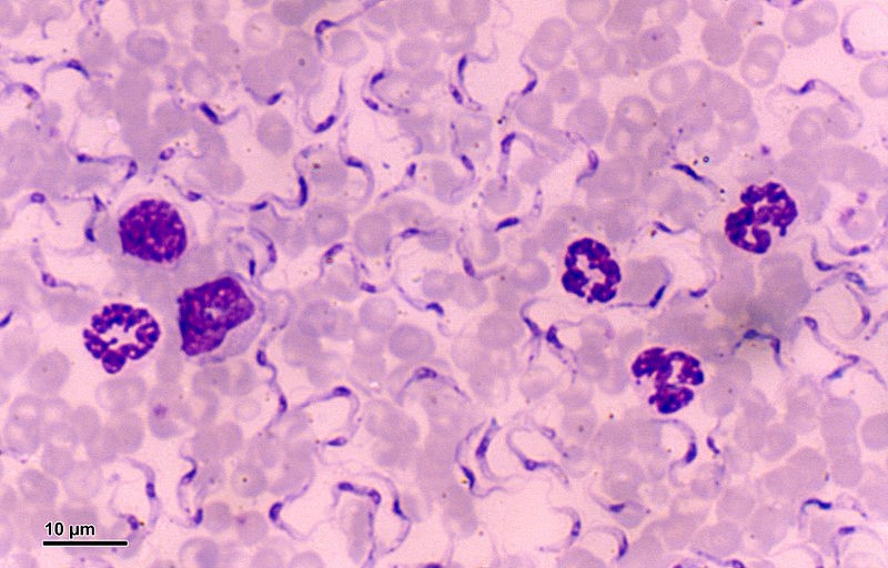
Trypanosoma equiperdum under microscope
ETIOLOGY
Dourine or Dollars disease is a parasitic venereal disease whose causative agent is a flagellate protozoan i.e. Trypanosoma equiperdum which belongs to order Trypanosomatida family Trypanosomatidae and subgenus Trypanozoon.
EPIDEMEOLOGY

Distribution of dourine in different parts of the world (Yonas 2015)
The disease is transmitted during sexual intercourse or in some cases infected mares pass infection to foals. Invertebrate vector is not involved in case of dourine. Dourine is endemic in regions of Africa, Asia and Latin America as well as Middle east and Western Europe.
HOST- It includes animals of Equidae family i.e. horses, donkeys, and mules. Infected animals are generally the reservoirs of the disease.
TRANSMISSION– Generally transmitted during coitus mainly from stallion to mare but vice versa is also possible. Rarely it is possible that foals may get infection via mucosa during birth or through milk.
SOURCES OF INFECTION-Vaginal secretions of infected mares, seminal fluid, mucous exudate of penis, mammary secretions of some animals.
CLINICAL SIGNS
The clinical signs depend on virulence of strain, nutritional factors, and stress factors.
Small amount of vaginal discharge is initial sign in case of mares.
Vaginal discharge
- Swelling and oedema of vulva extending up to udder and ventral abdomen in later stages.
- Vulvitis, Vaginitis along with polyuria.
- Significant abortion in case of virulent strains.
- Initial signs in stallions include oedema of prepuce and glans penis extending up to scrotum ventral abdomen and thorax.
- Vesicles and ulcers on genitalia which leave behind permanent white scars called as leukodermic patches.
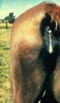
- Circular cutaneous plaques in some locations sometimes called Silver Dollar Plaques-Pathognomonic sign, hence the disease is also called as Dollar’s disease.
Typical silver dollar oedematous skin plague (arrowed)on hind leg of horse infected with dourine. Image courtesy of the Istituto G. Caporale -Teramo, Italy
- Other signs may include restlessness, shifting weight to one leg, anaemia, conjunctivitis, keratitis, recumbency and paralysis.
LESIONS
Gross Pathological Lesions
Cachexia, Anemia, Muscular Hypotrophy, Genital lesions, Cutaneous oedematous plaques, gelatinous exudates present under skin ,testicular tunica and scrotal sheath infiltrated in case of stallion ,Gelatinous infiltration of vaginal mucosa , uterus, vulva, mammary glands etc. in case of mares.
Microscopic Lesions
- Hemosiderin deposition in spleen.
- Iliac, supramammary and popliteal lymph nodes show non specific reactivity with hyperplasia of plasma cells which is a sign of increased haemolymphatic activity.
- Oedematous plague with pustular dermatitis around lesion along with vacuolar degeneration.
- Neurodegenerative lesions and inflammatory vasculitis of CNS with oedematous infiltration.
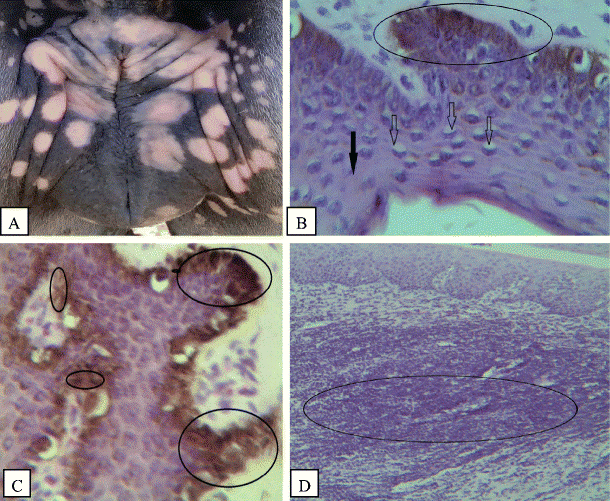
- A)Depigmentation of the vulval lip (gross). B)Vacuolar degeneration of the cells (lighter arrows) and necrotized cell (darker arrow) in the stratum spinosum, degeneration, and necrosis of the basal cells with melanin pigment were evident (circled areas). C) Excess free melanin in the stratum spinosum (small circles) and within the basal layer (large circles). D) Severe dermatitis with infiltration of lymphocytes and plasma cells in the epidermis and dermis (circled areas) (Yonas 2015)
DIAGNOSIS
- There are different methods for diagnosis of Trypanosoma equiperdum. However, diagnosis in horses which are in chronic condition is difficult because parasites are not easily found or visible in tissues and bloodstream. Practically diagnosis is based on clinical evidence supported by serology.
- The Compliment Fixation Test (CFT) is the test prescribed by International Animal Health Code for testing equines before their transportation between countries.
- ELISA protocols were developed in past and compared with other serological tests.
- Blood samples are also examined by light microscopy and PCR for trypanosomes. `

The swelling of the genital organ of the dourine-infected stallion and sampling of trypanosomes from the urethral tract. a and b the swelling of the genital organ of the dourine-infected stallion. c HMI-9 was injected into the urethral tract using a transfer pipette to detach adherent T. equiperdum from the urethral tract mucosa. d Sampling of T. equiperdum from the urethral tract mucosa using a cotton swab
Different diagnosis method include:
Observation of clinical manifestations and lesions resembling dollar spots can be complemented by diagnostic methods such as Serological tests like CFT (Complement Fixation Test), IFAT (Immuno Fluorescent Antibody Test), ELISA (Enzyme Linked Immunosorbent Assay), and Immunochromatography. Additionally, the microscopic examination of fresh fluids, such as mammary secretions and vaginal exudate, serves as a valuable diagnostic approach. Furthermore, the isolation of the parasite is a crucial step in the diagnostic process.
TREATMENT
There are no officially approved drugs used to treat dourine. However, evidence from in vitro drug sensitivity of T. equiperdum indicate that Suramin, Diminazine, Quinapyramine, and Cymelarsan are effective against it.
- Cymelarsan @0.25-0.5mg/kg body weight is found effective.
- Suramine at 10mg/kg body weight IV
- Quinapyramine dimethylsulfate at 3-5mg/kg SC
PREVENTION AND CONTROL
- Prevention of natural mating and AI with infected horses or semen.
- Testing of blood for presence of antibodies against equiperdum.
- Test and slaughter policy.
- Movement control influenced by legislation.
- Introduction of horses from endemic regions should be avoided and if done then it should be after proper quarantine and testing of animal.
- Proper implementation and monitoring of policies is equally essential to avoid chances of outbreak and thus prevent the disease.
REFERENCES
- Foreign Animal Diseases ,7th edition, Revised 2008, Committee on Foreign and Emerging diseases of the United Staes Animal Health Association, Dourine pg.231-236.
- https://en.wikipedia.org
- Clausen, P. H., Chuluun, S., Sodnomdarjaa, R., Greiner, M., Noeckler, K., Staak, C., … & Schein, E. (2003). A field study to estimate the prevalence of Trypanosoma equiperdum in Mongolian horses. Veterinary Parasitology, 115(1), 9-18.
- Gizaw, Y., Megersa, M., & Fayera, T. (2017). Dourine: a neglected disease of equids. Tropical animal health and production, 49, 887-897.
- Brun, R., Hecker, H., & Lun, Z. R. (1998). Trypanosoma evansi and T. equiperdum: distribution, biology, treatment and phylogenetic relationship (a review). Veterinary parasitology, 79(2), 95-107.
- Pascucci, I., Di Provvido, A., Cammà, C., Di Francesco, G., Calistri, P., Tittarelli, M., … & Caporale, V. (2013). Diagnosis of dourine in outbreaks in Italy. Veterinary Parasitology, 193(1-3), 30-38.
- Suganuma, K., Narantsatsral, S., Battur, B., Yamasaki, S., Otgonsuren, D., Musinguzi, S. P., … & Inoue, N. (2016). Isolation, cultivation and molecular characterization of a new Trypanosoma equiperdum strain in Mongolia. Parasites & vectors, 9(1), 1-9.
- Vulpiani, M. P., Carvelli, A., Giansante, D., Iannino, F., Paganico, D., & Ferri, N. (2013). Reemergence of dourine in Italy: clinical cases in some positive horses. Journal of Equine Veterinary Science, 33(6), 468-474.