Examination of Milk: A New Approach of Handling Bovine Mastitis
Rajeev Ranjan1, Jitendra Kumar Biswal1 and Madhurendu Kumar Gupta2
1Scientist, ICAR-Directorate of FMD, Mukteshwar-263138, Nainital, Uttarakhand
2Professor and Head, Department of Veterinary Pathology, Ranchi Veterinary College, Ranchi, Jharkhand
Bovine mastitis is a highly prevalent disease in dairy cattle, and one of the most important diseases affecting the world’s dairy industry; it places a heavy economic burden on milk producers all over the world. Mastitis is inflammation of parenchyma of mammary gland characterized by physical, chemical and usually bacteriological changes in milk and pathological changes in glandular tissues. Mastitis thus has become major area of concern in the field of veterinary clinical practice. Despite the voluminous scientific work and literature published on mastitis, the problem still persist owing to lack of scientific approach in the diagnoses and treatment of bovine mastitis. The situation has been complicated by the continued indiscriminate use of antibiotics, wrong approach of selection of suitable antibiotics after culture and sensitivity test of milk. However it is difficult to judge the clinical efficacy of a drug solely on in vitro test, as there is large variation in response among herds and within herds owing to type of organism involved, degree of udder induration, physico-chemical properties and kinetic behavior of drugs in udder & milk, site of injection and sensitivity of udder pathogens. The in vitro efficacy of an antibiotic against the isolate may not be achieved in vivo since many factors affect the penetration of drug into the udder tissue/site of infection like lipid solubility and tissue protein binding of the drug, pH of milk and inflammatory exudates/ barrier at the site. Thus, following culture and sensitivity test the additional information on factors like presence of fat globules, reticulin, pH of milk sample etc. could prove to be of vital importance in selection of medicines.
Economic losses: Mastitis is the major cause of economic loss to dairy industry globally. Annual losses in the dairy industry due mastitis was approximately 2 billion dollar in USA and 526 million dollars in India, in subclinical mastitis and clinical mastitis are responsible for approximately 70% and 30% respectively of dollars losses. Mastitis accounted for 26% of the total cost of all dairy cattle diseases. Losses from mastitis were twice as high as losses from infertility and reproductive diseases. The economic losses sustained by dairyman or farmers due to mastitis has been identified likes reduced milk production, discarded milk, early cow replacement costs, reduced cow sale value, drugs, veterinary services, labor etc.
Etiology: Mastitis, an inflammation of the mammary gland, is a complex of polymicrobial origin. Mastitis-causing pathogens includes bacteria likes Staphylococcus aureus, coagulase negative Staphylococcus spp., Streptococcus spp., E. coli, Pseudomonas spp., Klebseilla spp., Bacillus spp., and non-bacterial pathogens, likes mycoplasms, fungi, yeasts, and chlamydia. These pathogens infect the udder generally through the ducts papillary, which is the only opening of the udder to the outside world. Despite intensive etiological research, still around 20–35% of clinical cases of bovine mastitis have an unknown aetiology. Miltenburg (1996) found a 28% negative rate in 1045 cases of clinical mastitis, and Wedderkopp (1997) did not note pathogens in 35% of 6809 milk quarters in 3783 cows suffering from clinical mastitis and Ranjan (2010) has also reported there is no isolate in 52 cases of clinical cases of bovine mastitis out of 190 positive samples of bovine mastitis. An explanation for these high percentages of culture-negative samples might be a low concentration of udder pathogens. In some form of mastitis, caused by E. coli or other environmental organisms, systemic effects of endotoxin continue in the absence of viable bacteria in milk. Some of the microorganism such as Mycoplasma spp, Listeria spp. etc require specialized isolation media, no growth may also be observed in traumatic mastitis. In addition, the reason behind no isolation of bacteria from milk samples may include treatment with antibiotics before sampling and elimination of the bacteria in the course of inflammatory reaction. Lack of bacterial growth may also be a feature of chronic mastitis in which the organisms have been eliminated but the pathological changes persist. But these agents cannot be the explanation for all culture-negative milk samples from mastitis cows, because these agents are no common udder pathogens.
Transmission of bovine mastitis: It is mainly transmitted by contact with contaminated milking machine, and through contaminated milker hands or materials.
Types of bovine mastitis: There are two type of bovine mastitis i.e. clinical and sub-clinical bovine mastitis.
- Clinical bovine mastitis: Clinical mastitis may be categorized in to different types: per-acute, acute, sub-acute and chronic.
- Per-acute Mastitis: sudden onset, severe inflammation of the udder, and serous milk-Systemic illness often precedes the symptoms manifested in the milk and mammary gland.
- Acute Mastitis: sudden onset, moderate to severe inflammation of the udder, decreased production, and occurrence of serous milk/fibrin clots, Systemic signs are similar but less severe than for the per-acute form.
- Sub-acute Mastitis: Mild inflammation, no visible changes in udder, but there generally is small flakes or clots in the milk, and the milk may have an off-color. There are no systemic signs of illness.
- Chronic Mastitis: Chronic mastitis may persist in a subclinical form for months or years with occasional clinical flare-ups. Treatment usually involves treating the clinical flare-ups, or culling the cow from the herd.
- Sub-clinical bovine mastitis: The most common form of mastitis, 15×40 X more common than clinical mastitis, no gross inflammation of the udder and no gross changes in the milk, decreased production and decreased milk quality. In this mastitis only there is increase in the number of Somatic Cell Count.
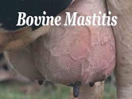
Clinical signs and symptom: Disease can be identified by abnormal changes in the udder likes swelling, heat, redness, hardness or pain and change in the colour and consistency of milk such as a watery appearance, flakes, clots, or pus, red, thick yellow, thick green etc (Fig-1).
Changes in the composition of mastitis milk: Due mastitis there are change in composition of milk and leads to decline in potassium, lactoferrin, casein, the major protein in milk and calcium level.
Diagnosis: Diagnosis of bovine mastitis is done on the following conditions:
- Physical examination of udder: Udder becomes hot, swollen, pain, red.
- Tests of milk: Abnormality of milk can be done by the strip cup test, bromothymol blue test, chloride test, hotis test, California mastitis test, white side test, catalase test etc.
- Direct test: In this test there is isolation and identification of the organisms from the suspected milk sample.
Examination of milk: Like blood, fecal and urine examination tell us about diagnosis and that help in treatment of etiology. Same if we follow the report of milk examination then it will help in treatment of bovine mastitis. Report of milk examination contain following point:
- Physicochemical examination of milk: In this examination there is recording of colour of milk, consistency of milk, odour and pH of milk.
- Microscopic examination of milk includes somatic cell counts (lacs/ml), clumping of somatic cell, lipid vacuole (fat droplets), fibrin/reticulin, differential leukocyte count and RBC.
- Bacteriological study includes microorganism isolated and colony count of organism.
- Antibiotic sensitivity test includes list of antibiotics sensitive and resistance against isolated organisms.
Although the changes in consistency of milk due to different isolate were overlapping in nature, the occurrence of thick and purulent consistency was observed mainly in pseudomonas, staphylococcal and yeast mastitis. The normal cow milk pH ranges between 6.4 – 6.8. Highest rise of pH was recorded in cases of staphylococcal mastitis particularly during Staphylococcus aureus infection. Since blood contains nearly four times the concentration of sodium and chloride in milk; condition like mastitis, which brings alteration in the permeability of the mammary gland vasculature, will raise the salt concentration of milk. Increasing permeability is also accompanied by diffusion of alkaline blood components, principally bicarbonate into milk thus raising the pH of milk. Therefore, higher pH of milk sample in cases of staphylococcal mastitis and less commonly streptococcal, E. coli and pseudomonas mastitis suggests that these agents might increase the mammary gland vasculature’s permeability during mastitis. It demands that the drugs acting at higher pH like amikacin, streptomycin, gentamicin and lincomycin should be preferred, if the milk samples are showing high pH.
Clumping of somatic cells in the milk smear was a feature found in mastitis. It was most prominent feature of mastitis caused by E. coli followed by coagulase negative Staphylococcus spp., Staphylococcus aureus and Pseudomonas. In mastitis caused by Staphylococcus aureus, the enzyme coagulase could play an important role in clumping. This fibrinogen binding protein on the staphylococcal cell surface can directly convert fibrinogen to insoluble fibrin. Clumping factor (coagulase) had been reported to be synthesized in greater amount during chronic infection, which implies that clumping of somatic cells is a good indicator for detecting chronicity of mastitis on the basis of exfoliative cytology.
Early detection of reticulin fibre in the milk by silver impregnation technique, thus could serve as an excellent marker for detection of onset of fibrosis in the udder. The reticulin condenses to form collagen fibres, which under the influence of remodeling enzymes metalloproteases under go bonding and weaving to form permanent scar tissue. Additionally, the information could be utilized for selection of antifibrotic therapeutic agents such as hylase, streptokinase, streptodornase and hyalurinidase, which could be used in conjunction with antibacterial agents.
Papanicolaou staining revealed presence of lipid vacuoles in mastitis milk. Lipid globules play major role in determining effectiveness of phagocytic capability of neutrophils. These when phagocytosed by the neutrophils result into reduced efficacy of neutrophils as a bactericidal cell population because of loss of myeloperoxidase enzyme and other hydrolytic enzymes in response to phagocytosis of fat globules. It may also be noted that drugs such as chloramphenicol, ciprofloxacin, enrofloxacin and pefloxacin having high lipid solubility and ability to cross the biological barrier could prove to be more effective in lipid rich milk as detected during cytological study.
After completion of physicochemical and microscopic examination of milk there is need of isolation of microorganisms and antibiotic sensitivity test. Once the antibiotic sensitivity test completed make a list of sensitive and resistance antibiotics against isolated organisms. Inclusion of the parameters in the routine laboratory testing of the milk samples of bovine mastitis like blood, fecal and urine examination will be of definite help in assessing the severity of infection, presence of causative agents in the milk sample and selection of therapeutic regimen as well as assessment of the duration of treatment.
Thus, it could be concluded that the case of bovine mastitis should be handled carefully and reported comprehensively (Appendix-A) with respect to its diagnosis through conveniently performed parameters under field conditions like microscopic examination of milk with special emphasis on milk pH, clumping of cell population, lipid vacuolation and presence of reticulin. These preliminary tests should be followed by bacteriological examination and antibiotic sensitivity testing for judicious selection of drug in order to achieve maximum containment of bovine mastitis in field condition. Inclusion of the parameters in the routine laboratory testing of the milk samples of bovine mastitis will be of definite help in assessing the severity of infection, presence of causative agents in the milk sample and selection of therapeutic regimen as well as assessment of the duration of treatment. Thus, the present study has got definite applied value.
Summary: Bovine mastitis is a highly prevalent disease in dairy cattle, and one of the most important diseases affecting the world’s dairy industry. Mastitis is the major cause of economic loss to dairy industry globally. Annual losses in the dairy industry due mastitis was approximately 526 million dollars in India. Despite the voluminous scientific work and literature published on mastitis, the problem still persists owing to lack of scientific approach in the diagnoses and treatment of bovine mastitis. The situation has been complicated by the continued indiscriminate use of antibiotics, wrong approach of selection of suitable antibiotics after culture and sensitivity test of milk. However, it is difficult to decide the clinical efficacy of a drug only on in vitro test, as there is large variation in response among herds and within herds. The in vitro efficacy of an antibiotic against the isolate may not be achieved in vivo since many factors affect the penetration of drug into the udder tissue/site of infection like lipid solubility and tissue protein binding of the drug, pH of milk and inflammatory exudates/ barrier at the site. Thus, following culture and sensitivity test the additional information on factors like presence of fat globules, reticulin, pH of milk sample etc. could prove to be of vital importance in selection of medicines. The present study suggested that comprehensive examination of milk and antibiotic sensitivity testing could provide valuable information with respect to nature of disease, and selection of drug regimen for the prevention and treatment of mastitis.
APPENDIX- A
THE REPORT ON EXAMINATION OF MILK |
||||||||||
| A. Physicochemical Examination | : | |||||||||
|
|
a
b c d |
Colour of milk | : | |||||||
| Consistency of milk | : | Normal/ Serous/ Purulent | ||||||||
| Odour | : | Normal/ Offensive | ||||||||
| pH of milk | : | |||||||||
|
B. Microscopic Examination |
: |
|||||||||
| a
b
c
d
e
f |
Somatic cell count (lacs/ml) | : | ||||||||
| Clumping of somatic cell: | : | Present | Absent | |||||||
| Degree of clumping | ||||||||||
| Lipid vacuolation | : | Present | Absent | |||||||
| Degree of vacuolation | ||||||||||
| Fibrin/Reticulin | : | Present | Absent | |||||||
| Degree of reticulin | ||||||||||
DLC |
: | Neutrophil | …….. % | |||||||
| Lymphocyte | …….. % | |||||||||
| Monocyte | …….. % | |||||||||
| RBC | : | Present/Absent | ||||||||
|
C. Bacteriological study |
: |
|||||||||
| a
b |
Microorganism isolated | : | ||||||||
| Colony count: | : | |||||||||
|
D. Antibiotic sensitivity test |
: |
|||||||||
|
|
Sensitive to | Resistant to | ||||||||
| Signature
|
||||||||||
| NOTE: | SCC of >5 lacs/ ml is suggestive of infections. | |||||||||
| Qualitative gradation of microscopic examination is as follows: | ||||||||||
| Mild | + | |||||||||
| Moderate | ++ | |||||||||
| Marked | +++ | |||||||||
| Severe | ++++ | |||||||||


