First Aid Programme and Emergencies in Veterinary Practice
Dr Md Moin Ansari
Associate Professor/Senior Scientist
Sher-e-Kashmir University of Agricultural Sciences & Technology of Kashmir,
Faculty of Veterinary Sciences and Animal Husbandry
Division of Veterinary Surgery and Radiology,
Srinagar-190006, Jammu & Kashmir.
Abstract:
First aid is warranted in situations of life-threatening emergencies which require immediate action by the owner or animal health workers. In some cases, first aid consists of the initial support provided to animals in the middle of a medical emergency. It is important for owners of animals to be aware of this potentially life-threatening health condition. If you suspect your animal is showing signs of illness/injury, contact your veterinarian immediately. Do not apply materials (other than those mentioned) to the illness/injury unless specifically instructed to by your veterinarian. Timely, early intervention and proper first aid programme prevents the situation from deteriorating further, live saving, promotes recovery of the ailing animal and reduce economic losses to the owners.
Key words:
First aid, emergency, injury, illness, management
Introduction
First aid means immediate/basic veterinary care and treatment of injuries or any other sudden illness in the animals. Timely and proper first aid programme prevents the situation from deteriorating further and promotes recovery of the ailing animal. In some cases, first aid consists of the initial support provided to animals in the middle of a medical emergency. First aid is warranted in situations of life-threatening emergencies which require immediate action by the owner or animal health workers. There are three basic principles of C’s to remember—check, call, and care. When it comes to first aid, there are three basic P’s to remember—preserve life, prevent deterioration, and promote recovery. First aid is as easy as ABC – airway, breathing and CPR (cardiopulmonary resuscitation). Animal owners can provide significant medico-veterinary assistance at the scene of any injury or insult to the body parts. At the time of the initial telephone call, the animal owner should be questioned about the level of consciousness, breathing pattern, type of injury or toxicity, and even some aspects of the animal’s perfusion (viz: gum color, level of responsiveness). When the patient arrives at the veterinary hospital, regardless of the presenting complaint, a triage should be performed that assesses vital parameters, including: 1. temperature, 2. pulse rate, 3. respiratory rate, 4. level of consciousness and 5. level of pain. In addition to that assessment of perfusion can include pulse quality, mucous membrane color, and capillary refill time. An abnormality in any one of these or one of the presenting complaints listed below warrants urgent evaluation by a veterinary doctor. A first aider is the term describing any person who has received a certificate from an authorized training body indicating that he or she is qualified to render first aid.
Aims and Objectives of First aid
It is vital that the owner adequately restrain the pet before starting any first aid procedures to ensure the safety of the pet owner and pet. When moving the animal, motion of the head, neck, and spine should be minimized. A flat, firm board of wood, cardboard, or thick fabric can be used to provide support. Following are the aims and objectives of the first aid in veterinary practice:
- the first concern is for the safety of the animal owner.
- to save life of the animals.
- to reduce their pain and suffering.
- to minimize injury and future disability.
- to stay safe and calm at all times.
- to assess a situation quickly and calmly and summon the appropriate help if necessary.
- to assist the casualty and provide the necessary treatment, with the help of others if possible.
- to pass on relevant information to the emergency services, or to the person who takes responsibility for the casualty.
- timely first aid prevents the situation from deteriorating further and promotes recovery of the ailing animal.
- provided in case of infectious and noninfectious diseases, any sort of wounds, electrocution and burns, etc.
- prevention and control of transmission of diseases,
- preparation of common antiseptic solution,
- cleaning of muzzle and hooves and
- necessary items for a veterinary practice first aid kit.
First aid kit for veterinary practice:
As well as knowing some basic first aid programmes, it is important that households and workplaces have a first aid kit that meets their needs and is well organized, fully stocked and readily available at all times. The contents should be appropriate to cope with a range of emergency situations, depending on the setting. It’s a good idea to have a number of kits handy in different places, such as in the home, car or office. First of all, it is important to ensure that the first aid kit is easy to find and carry. The first aid kit must be kept clean and dry. Suggested list of items for the first aid kit:
- Allis Tissue Forceps: used for grasping slippery or dense tissue. 2. Crile Forceps used for: clamping blood vessels or tissue before cauterization or ligation. 3. Kelly Forceps (Curved and Straight): primarily used for clamping large blood vessels or manipulating heavy tissue. They may also be used for soft tissue dissection. 4. Sponge Forceps: used in surgical procedures to hold gauze Needle Holders: Small 5.5”, Medium 6.5”, Large 7.5”. 5. Olsen-Hegar Needle Holders built in scissors for easy access to cut suture. 6. Scalpel Blade Handle (No. 3 and No. 4): used for cutting surface tissue or sutures, both may be used interchangeably. 8. Scissors: Scissors have become a multipurpose instrument used in various surgeries Metzenbaum Scissors (Small 5.5”, Large 7.0”, Very Long, 9.0”): used for dissecting and cutting tissue. These scissors are not recommended for cutting sutures, drains, or heavy tissue. Tenotomy Scissors used for delicate dissection and cutting, in ophthalmologic surgeries. Thumb Forceps: These forceps have a wide, flat thumb grasp area that is commonly serrated. The jaws are short and the tips are narrow. Adson Dressing Forceps (“thumb” forceps): They have a wide thumb grasp for increased precision and control. These thumb forceps are straight with delicate, serrated tips. Adson Tissue Forceps, (small rat tooth) are for holding and manipulating delicate tissues. The jaws are short and the tips are narrow. Rat Tooth, larger rat tooth style forceps are commonly used for grasping fine tissue and blood vessels or soft tissue dissection. The narrow tips and atraumatic teeth cause little to no damage to the tissues. It is used for draping in towels during surgical procedures Direction Probe with eye (5 ½”). 10. Flashlight: used for illuminating a local area or cavity of the patient. 11. Halter and rope: A halter or headcollar is headgear that is used to lead or tie up livestock and, occasionally, other animals; it fits behind the ears (behind the poll), and around the muzzle. To handle the animal, usually a lead rope is attached. On smaller animals, such as dogs, a leash is attached to the halter. 12. Wire cutters. 13. Disposable gloves . 4×4 gauze sponges 15. Skin cleanser. 16. Light Cloth: Placing a light cloth over the head of the animal can lessen external stimuli that may cause fearful and aggressive reactions. 17. Owners can be instructed as to how to muzzle most dogs using a long strip of fabric if there are no facial injuries or respiratory distress. 18. Dark boxes: Cats can be placed in dark boxes to minimize stress during transport; the box should have holes large enough so that the cat can be observed and to allow adequate fresh air. 18. Several small bottles of sterile saline. 20. Rolls of medical tape. 21. Fly repellent (Himax ointment). 22. Several large syringes (5ml, 20 ml, 60 ml). 23. Cotton. 24. Antibiotic eye ointment (Ciprofloxacin, Gentamycin). 25. Digital thermometer
Procedure for First Aid:
- Owners should be asked whether haemorrhage is ongoing or whether bleeding was seen at the site of injury.
- To help control external bleeding, place a compress of clean cloth or gauze directly over your dog or cat’s wound. Apply firm but gentle pressure, and allow it to clot. If blood soaks through the compress, place a fresh compress on top of the old one and continue to apply firm but gentle pressure. Pulsating arterial bleeding should be controlled by direct digital pressure or a pressure bandage. Any long pieces of fabric or gauze can be used. Often, washcloths and hand towels are adequate when applied. Additional material can be placed over the original bandage if it becomes soaked with blood. If the bleeding from a limb is venous (dark, oozing), the limb can be elevated above the heart. Tourniquets should be used only on appendages (viz: limbs, tail) when compression wraps have failed to control bleeding. The tension on the tourniquet must be relaxed every 5–8 minutes to allow blood flow to the distal limb and then re-tightened after 2 minutes.
- Wound may be defined as disruption of the cellular or anatomical continuity of the normal organ structure, caused by physical, chemical, mechanical or biological agents. When the tissues damaged different types of microorganisms will enter the wound and in a number of ways (direct contact, airborne dispersal, self-contamination) which initiate wound contamination and the development of infection. In wound affections first step is to arresting the hemorrhage/bleeding and then cleaning and disinfection. Wound is covered with sterile gauze, surrounding area cleaned, hairs clipped, shaved and cleaned thoroughly with soap and water. Apply weak antiseptic solution (iodine solution). Prevention of further trauma and rest/immobilization: Give rest to the animal after proper bandaging, animal should be given rest and protect the affected part from licking, biting or rubbing etc. Despite some recent advances in understanding its basic principle, problem in wound healing continues to cause significant morbidity and mortality. There are three types of symptoms depends upon the degree of tissue destruction. There are a number of indicators of wound infection, these includes: localized erythema, localized pain, localized heat, cellulitis and edema. i. Local symptoms include haemorrhage/bleeding, pain and gaping of the lips of the wound. ii. General symptoms depend on the depth and site of the wound. These comprise of febrile disturbances, neuritis and paralysis when nerves are involved in the wound. Remote symptoms: is observed on a part away from the wound like abscess formation, in the neighbouring tissues due to infection through lymphatic channel. The lymph gland may show inflammation and abscess formation.
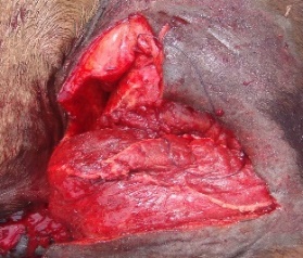
Laceration wound
Basic principle of wound treatment (in general): In wound management the Primary objective is to establish the condition and an environment which are essential for rapid and complete wound healing. Recent wounds require immediate emergency/first aid treatment to control hemorrhage and to protect it from further injury. Lavage the wound thoroughly and remove the foreign bodies and dirt particles including blood clots, tissue debris, and metallic objects by gauze soaked in sterile distilled water or normal saline solution. Measures should be taken to overcome the bacterial growth. This is done by antiseptic (Chlorhexidine diacetate (0.05%), Povidone iodine (0.1 or 1%), Tris EDTA, Acetic acid (0.25 or 0.5%) cleaning of the wound and by the use of antibiotics (penicillin, oxytetracyline, amoxicillin, enrofloxacin etc.). If the wound is less than 6-8 hours old, debridement is carried out. Provision for drainage: wound drainage is necessary, when pockets cavity is present. Drains are used for the maintenance of a passage way for the removal of accumulated pus, serum and clotted blood within the cavity or dead space. Closure of wound: accurate apposition of the wound surfaces influences the early healing of the wound. Questions of closing the wound gap must be taken on the basis of local condition of the wound. It the wound is fresh and of condition incised nature, the edges are aligned in correct anatomical relationship to take primary healing. In case of larger wound, suture is applied only when haemorrhage has stopped and further sloughing and bacterial invasion are unlikely so that primary healing may be expected. If there is any doubt, the lowest part should be kept open to provide drainage and to ensure healing. Prevention of further trauma and rest/immobilization: this is achieved by application of firm, well-padded immobilizing bandage. This is very essential for wound affecting the extremities. Proper bandage and immobility also prevent edema. Healing is rapid and scar is reduced.
- Penetrating foreign objects (viz: sticks, arrows) should be left in place during transport; however, care should be taken to guard against movement of the object to prevent further injury. It is often necessary to stabilize the shaft of the penetrating object just outside the body and, holding it firmly, cut off the shaft, leaving a portion exposed so it can be easily located.
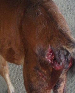
- Any bite wound, including a dog bite requires to be tended right away to reduce the risk of bacterial infection. The wound is assessed to determine the severity. In some instances, first aid can be applied at home however, in other cases immediate veterinary medical care may be required. Dog bite wound should be treated by clean the blood and apply an antibacterial ointment. If the wound is bleeding then apply and press a clean cloth to the area to stop bleeding. Clean the area and apply a sterile bandage. It is important to seek immediate medico veterinary attention in case of a bleeding dog bite wound.
Dog bite wound
- Snake bite should be treated with antivenin administered at the hospital by registered veterinary doctor is the most direct and helpful treatment for your pet and livestock. Ideally, Antivenin is administered within two hours of the bite to be the most effective.
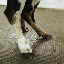
- First aid treatment for fracture includes stop any bleeding. Apply pressure to the wound with a sterile bandage, a clean cloth or a clean piece of clothing. Immobilize the injured area. Don’t try to realign the bone or push a bone that’s sticking out back in. Apply ice packs to limit swelling and help relieve pain. In animals with fractures below the elbow or stifle, support can be provided during transport. With any fracture, there is concern for additional damage to muscles, nerves, vessels, and bones as well as pain if support of the fracture is not provided. Once the animals have been adequately restrained, the owner can make a support splint from a rolled newspaper or magazine, which is secured in place by long pieces of fabric or duct tape. Fractures above the elbow or stifle are challenging to immobilize. Patient movement should always be minimized.
First aid in fracture management
- Animals with altered mentation after trauma should be transported with the head level with the body or elevated 20°C. There should not be any jerking or thrashing motions, and manipulations of the neck or occlusion of the jugular veins should be avoided.
- If a toxic ingestion is present, the animal owner should be instructed to bring the animal immediately to a veterinary doctor at hospital. If possible, the container of the toxin should be brought in for identification.
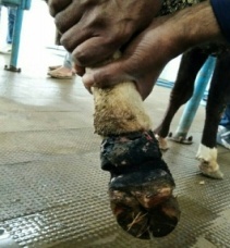
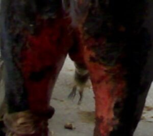
- Burns should be treated immediately with immersion in cool water or saline (salt and purified water) or spraying the affected area with cool water or saline. Obtain veterinary care quickly.
Chemical Burn in a bull
- Hypothermia should be managed by wrapping them up warmly in coats and blankets, and increase the room temperature if possible. Use body warmth and wrapped warm water bottles to gently warm them through. Get veterinary advice quickly.
- Dystocia, the difficulty in passing the fetus through the pelvic canal, is a common livestock emergency. Dystocia is always considered to be an emergency condition that demands immediate attention for the survival of both calf as well as dam. Get veterinary advice quickly. Positive clinical outcomes can be expected only when the clinician has a thorough understanding and knowledge of normal canine parturition, the pathogenesis and underlying etiology of dystocia, the criteria for diagnosing dystocia, and the appropriate medical and surgical interventions.
- One of the most common health emergencies in large and small ruminants is urolithiasis, or urinary stones, causing a blockage in the urinary tract. These blockages occur almost exclusively in males, especially castrated males. The urethra of females and intact males are usually larger, making blockages less likely. It is important for owners of large and small ruminants to be aware of this potentially life-threatening health condition. If you suspect your animal is showing signs of urinary blockage, contact your veterinarian immediately.
- In shock lie the animal on their right-hand side. Put a folded blanket under their lower back to raise it. This encourages blood to flow to their heart and brain. Get veterinary advice quickly.
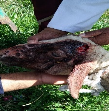
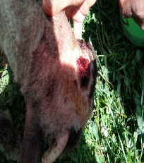
- Horn injury: depending on the extent of the horn injury, the horn may or may not grow back. Some horns injured at the base, or scurs that erupt from improper disbudding. It should be prepared to manage horn injuries, a first aid kit should include a blood stop agent, a wire saw, a sanding block, a cauterizing tool, gauze, wrap, antiseptics, antibiotics, pain management medication, and fly repellent. Get veterinary advice quickly.
Before treatment (Horn Injury) After the treatment (Horn Injury)
References:
Ansari MM 2014. Fundamentals of General Veterinary Surgery (A book for both undergraduate and postgraduate level). First edition, Published by Satish Serial Publishing House, Delhi-110033 (India). ISBN 978-93-81226-00-0. Pp. 1-408.
Dar, K.H., Ansari, M.M., Bhat, M.M. and Dar, D.H. (2014). Studies on dog bites wound of domestic animals and avian in Kashmir valley. International Journal of Veterinary Science. 3(3): 151-154.
Misk NA, Semika MA and Misk TN 2008. Predilection seats of body surface abscesses in relation to the way of infection in some domestic animals. 25th World Buiatrics Congress, Budapest, Hungary.
Peer, F.U. and Ansari, M.M. (2006). Management of BHC poisoning in crossbred calves- a case report. The Indian Journal of Veterinary Science and Biotechnology. 9 (3): 51-52.
Smith BP 1983. Diseases of the alimentary tract. In Large Animal Internal Medicine. 2nd edn. Philadelphia, Mosby. pp 858-860.
Valentine BA 2004. Neoplasia. In: Farm Animal Surgery. (Fubini SL, Ducharme NG, Eds.), Saunders. An Imprint of Elsevier, USA, pp.23-44.
https://www.pashudhanpraharee.com/first-aid-in-cattle-health-management-for-cattle-farmers/
***********************
https://veterinarypartner.vin.com/default.aspx?pid=19239&id=4951311


