HAEMORRHAGIC ABOMASITIS AND SEPTICEMIA – A CASE STUDY
D T Naik*, Rajendra kumar T2, Jyothi C3
- Professor and Head, Department of Veterinary Pathology
2) Assistant professor, Department of Veterinary Pathology
3) Assistant professor (contractual basis), Department of Veterinary Pathology
Department of Veterinary Pathology; Veterinary College, KVAFSU, Bidar, Karnataka.
E.mail: drdtnaik@gmail.com - ABSTRACT
- A carcass of two months old male goat was presented to the Department of Veterinary Pathology, Veterinary College, Bidar, Karnataka with a history of diarrhoea. The carcass was dehydrated. Visible mucous membranes were pale and the subcutaneous tissue was pale.
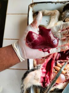
- The liver was enlarged and congested (Fig.1).
- Gall bladder was distended. Spleen was greately atrophied.
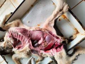
- The mesentery was severely congested (Fig.2).
- Mesenteric lymphnodes were enlarged and oedematous.
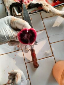
- Extensive haemorrhages over not separating the renal cortex and medulla along with great reduction in the size of renal pelvis (Fig.3).
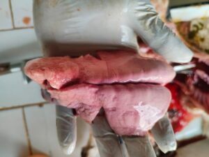
- Intestinal mucosa showed haemorrhagic enteritis. Heart was congested and lumen showed little currant jelly clot. Both the lungs were pale with slight congestion on the right apical lobe (Fig.4).
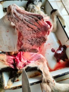
- Tracheal mucosa was slightly congested. The remarkable changes occur in the abomasums were resembling zebra markings in the mucosa (Fig.5).
- The cause of death was attributed to Haemorrhagic abomasitis and septicemic lesions with a doubtful case of PPR.
Fig.1 The liver was enlarged and congested Fig: 2 The mesentery was severely congested


