HYPERKERATOSIS OR TEAT-END CALLOSITY OF UDDER IN DAIRY CATTLE
Compiled & Edited by-DR RAJESH KUMAR SINGH, JAMSHEDPUR, 9431309542,rajeshsinghvet@gmail.com
.
Teat-end hyperkeratosis is a thickening of the skin that lines the teat canal and surrounds the external teat orifice. The condition is variously described as teat rings, teat flowers, teat erosion, callus formation, callosity, cornification or teat-end roughness. Histological examination of teat sections confirms that teat-end hyperkeratosis results from a localised hyperplasia of the Stratum corneum or Stratum corneum and Stratum granulosum skin layers .Therefore, descriptions such as “teat inversion”, “teat eversion” or “prolapsed teats” are incorrect.
Hyperkeratosis means “excessive keratin growth”. It is a normal physiological response to the forces applied to the teat skin during milking, either by a milking machine, a hand-milker or a calf. The onset and severity of hyperkeratosis is profoundly influenced by the over-riding effects of climate, seasonal and environmental conditions, milking management, herd milk production level and genetics of individual cows. Reports of teat-end hyperkeratosis problems are far more prevalent in high-producing herds, for example, and especially during the colder periods of the year. Despite the fact that the same cows are milked with the same equipment by the same operators throughout the year, milking machines or hand milkers often get the blame for poorer teat condition during the winter months.
Teat-end callosity
After repeated milkings, changes appear in teat-end tissue, resulting in the development of a callous ring around the teat orifice. Cow factors like teat-end shape, teat position, teat length, milk production, lactation stage, and parity show a relationship with callused teat-ends .As early as 1942, “eroded” teat orifices were linked to machine milking . It is clear from more recent histological studies that the observed changes result from an increase or build up of callous tissue around the orifice rather than an ‘erosion’ of teat tissue or the orifice. The changes are associated with mechanical forces exerted by vacuum and the moving liner during machine milking. The magnitude of the force depends on milking vacuum, pulsation vacuum, machine-on time, liner type, and teat shape .The huge variation in the frequency of callosity between herds using similar milking systems suggests that a major genetic influence to susceptibility should not be overlooked
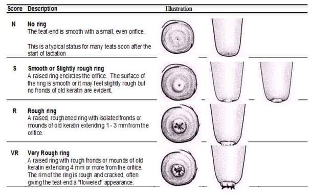
Teat-end callosity can be classified visually. Several systems have been developed (for example Sieber and Farnsworth, 1981, and more recently Shearn and Hillerton, 1996). The classification system adopted in The Netherlands includes marked differences in the thickness of the callosity ring (TECT), which is transformed to five classes: none (N), slight (A), moderate (B), thick (C) and extreme (D). Average TECT of teats was calculated by using the unit scores from 1 to 5. Additionally the ring is classified as smooth (1) or rough (2) (Neijenhuis et al., 2000). This system is proposed by the “Teat Club International” for research purposes
There are numerous factors which can influence the condition of the teats of a dairy cow. When farmers discuss teat condition, they are invariably describing teat end hyperkeratosis, an example of which can be seen in Fig. 1.
However, there are many other teat lesions and teat changes which can be measured and monitored which will provide an essential insight into what is happening during the milking process.
Some of the other important changes which can be measured would include:
o Post milking teat colour
o Firmness of the teat barrel
o Openness of the teat orifice
o Tissue ringing at the base of the teat
o Dryness of the teat
Discoloured teats after milking are highlighted in Fig. 2.
When changes occur to a cow’s teats after milking, these changes can be considered to fall into three categories. These changes are either:
o Short term changes (seen after one milking but soon disappear) such as colour of the teat, swelling and firmness of the teat end and teat barrel, palpable ringing of teat tissue where the teat attaches to the udder and the degree of openness of the teat orifice.
o Medium term changes (usually take a few days to develop) include changes in the teat skin condition and the incidence of petechial haemorrhages (blood blisters).
o Long term changes (normally take a number of weeks to develop) include the amount of teat orifice hyperkeratosis. However, when the environment is particularly harsh and the weather particularly cold or windy, hyperkeratosis can develop more rapidly.
The role of teat lesions——–
The teat canal is the primary physical barrier to invasion of pathogens into the udder. The smooth muscles which surround the teat canal should be contracted and the teat canal tightly closed between milkings to prevent pathogens entering the teat canal and then the udder.
This defence is aided by mature, lipid rich keratin cells lining the teat canal. In Fig. 3, the keratin lining of the teat canal has been stained red, and shows the convoluted nature of the canal. The keratin traps mastitis pathogens as they attempt to pass through the canal and these pathogens are then naturally removed from the udder during the milking process, when the shearing effect of milk flowing through the canal removes the outer layer of keratin.
When the teat end is in good condition and not rough or damaged and the skin of the teat is soft and supple, the teat is best placed to provide a natural barrier to the invasion of mastitis causing pathogens.
Any short term challenges to a teat can result in a reduction in the teats natural ability to resist bacterial challenge. While most attention is focused on teat end hyperkeratosis, the presence of other short term teat conditions such as discolouration, oedema, teat end wedging and congestion are less well recognised. However, studies in Holland have confirmed any form of circulatory impairment can be associated with an increase in the risk of sub-clinical mastitis infection.
When teat skin becomes dry, as well as often leading to an increase in teat end hyperkeratosis, the teat barrel becomes harder to clean. This can lead to an increased risk of infections caused by environmental mastitis pathogens such as Strep. uberis. Any open lesions on the teat skin can harbour contagious pathogens such as Staph. aureus. These lesions also cause discomfort for the cow during milk harvesting and can result in poor milking out. Fig. 4 illustrates a dry teat which will be difficult to clean.
Potential causes of adverse teat conditions
There are numerous factors which can affect the amount of teat end hyperkeratosis recorded including teat end shape, milk yield, peak milk flow rate, duration of milking and overmilking, stage of lactation, parity, teat skin condition and the interaction between the milking routine and the milking machine. The total time per day where the milk flow rate is less than 1.0 kg/min appears to have a significant effect on the level of hyperkeratosis found.
Cause of Hyperkeratosis
Major factors that can affect hyperkeratosis include teat-end shape, milk yield, peak milk flow rate, duration of milking and over-milking, stage of lactation, parity and the interaction between milking management and the milking machine. The total time per day when the milk flow rate is less than 1.0 kg/min appears to have a profound effect on the level of hyperkeratosis found.
In general, hyperkeratosis is more severe with long, pointed teats, slow milking cows and higher producing cows. Teat scores peak 3-4 months postpartum and decline as the lactation progresses.
The huge variation in levels of hyperkeratosis between herds employing similar milking systems with comparable levels of yield suggests that there may be a considerable genetic influence which should not be overlooked.

Reducing Hyperkeratosis
Although some hyperkeratosis is an obvious and probably natural response to milking, there are steps which can be taken to reduce the condition. It should be noted there is also some natural resolution depending on stage of lactation.
There is evidence that the total period of time when milk flow is less than 1 kg/min can influence hyperkeratosis levels. This period includes the time before the onset of milk let down, as well as the more commonly recognized period of time after completion of milking. It is perfectly possible to over-milk a cow at the start of the milking session if she is poorly prepared.
Cows which are thoroughly prepared prior to cluster attachment exhibit improved milk let down, which leads to shorter and more complete milkings with a clearly defined end of milk flow. Advances have also been made in adjusting detachment flow rates for automatic takeoffs (ATOs).
Ensuring the teat skin is in good condition, maintaining skin moisture and natural elasticity, will help the teat to restrict the development of hyperkeratosis. Using a high quality teat disinfectant, carefully applied, is essential.
There is also a need for a close examination of the genetic effect on hyperkeratosis. In addition to the obvious link with teat length and shape, there would appear to be a more subtle effect, which may in part be related to the type of teat duct keratin and the ability of certain teats to retain keratin.
Hyperkeratosis in dairy cows is a multi-factorial problem. However, it is easily quantified and could be suggested as a measure to assess the quality of management on a dairy herd and the perspective the herd owner has on the welfare of the dairy cow.
A small amount of teat-end callosity does not appear to increase the risk of intra-mammary infection in the lactating dairy cow, and may be considered as a beneficial physiological response of the teat to machine milking. A greater degree of teat-end callosity and roughness is associated with an increased probability of new intra-mammary infections. Evaluation of teat-end callosity in commercial herds may help to identify or resolve problems related to milking management, environment or the milking machine.
Reference-On Request


