INCLUSION BODY HEPATITIS(IBH)/ HYDROPERICARDIUM-HEPATITIS SYNDROM(HHS)/ANGARA DISEASE/LEECHI DISEASE
Dr Surinder Khanna, poultry consultant.
Hydropericardium syndrome (HPS) also known as hydropericardium–hepatitis syndrome/Angara disease (in Pakistan)/Litchi heart disease (in India) or inclusion body hepatitis–hydropericardium syndrome (IBH–HPS), is an emerging poultry disease in recent times characterized by sudde onset with a high death rate (Memon et al., 2006). The disease was named so due to accumulation of fluid in pericardial sac. The disease was first described in young broilers in Angara Goth, Pakistan, during the year 1987. Thereafter, it spread throughout Pakistan (Anjum et al., 1989) as well as neighboring countries like India (Dahiya et al., 2001). Currently, HPS is considered as an economically important endemic disease that can cause huge economic loss to poultry sector, especially in the South Asian region, owing to mortality, reduced productivity and immunodepression (McFerran and Adair, 2003).
The HPS was called as “Leechi” disease in India as the heart of the affected birds surrounded by hydropericardium resembled a peeled Indian Leechi fruit. The disease was first detected in Jammu and then in Punjab and Delhi in 1994 (Gowda and Satyanarayana 1994). Subsequently, the disease was continuously recorded with an average mortality of 34.6 % in broilers in Uttar Pradesh,Bihar and Haryana.
INTRODUCTION
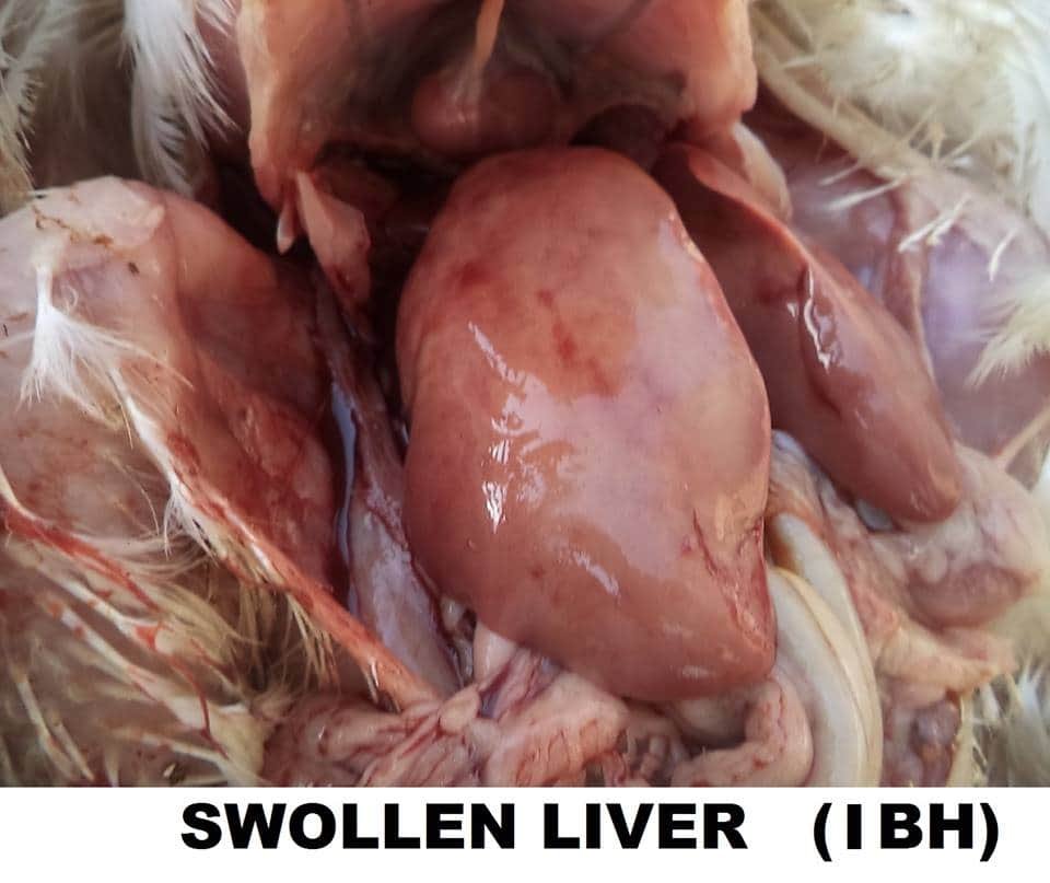
Adenoviruses are widespread throughout all avian species. Studies have demonstrated the presence of antibodies in healthy poultry and viruses have been isolated from normal birds. Despite their widespread distribution most adenoviruses cause no or only mild disease however some are associated with specific clinical conditions IBH outbreaks are encountered primarily in meat type chickens most commonly at the age of 3 – 8 weeks IBH often occurs as a secondary infection to immunodeficiency resulting from other diseases (IBD). On the background of dystrophic liver changes, hemorrhages of various intensity and size are outlines, thus creating a variety of liver lesions. The viral inclusion body hepatitis (IBD) is an avian adenovirus infection characterized by hemorrhages and dystrophic neurobiotic changes in the liver and kidney accompanied by intranuclear inclusion bodies. A characteristic macroscopic lesion is the enlarged, dystrophic liver with yellowish color and crumbly texture IBH is charaterised by acute mortality, often with severe anaemia, caused by an adenovirus a number of different serotypes have been isolated from disease outbreaks but they may also be isolated from healthy chickens this is also known as Litchi disease the virus is generally resistant to disinfectionts (ether, chloroform ph) and high temperatures.
Throught IBH outbreaks several serotypes from the 12 known avian adenoviruses (AAVS) of group are the sick chickens carry the virus in their excreta, kidneys, tracheal and nasal mucosa. The virus is resistant to many environmental factors and could be easily transmitted by a mechanical route. The transmission of adenoviruses is realized vertically by breeder eggs and horizontally, via excreta (mainly faces). In a number of cases the dominating lesion is the massive mouled or striated haemorrhages of the liver.
IBH is characterized by a sudden onset and a sharply increased death rate that reaches peak values by the 3rd and 4th day and returns back within the normal range by the 6th – 7th day. The total death rate is usually under 10% but sometimes could attain 30% more rarely macro scopically visible necrotic foci could be detected in the liver.
Cause —————
The disease is caused by a virus belonging to the group 1 of Avian Adenovirus (for example the Tipton strain) and is usually simultaneously accompanied by other immunosuppressive diseases such as infectious bursal disease or infectious anaemia. There are12 known serotypes of Avian Adenoviruses that may be involved in the development of this disease.
Transmission-——–
Egg transmission is an important factor but also horizontal transmission from bird to bird by contact with droppings can occur. Once the bird becomes immune, the virus can no longer be isolated from the droppings. Progeny of a shedding breeder flock can infect naive progeny of other breeder sources placed in the same house.
Diagnosis –———
Virus isolation and identification accompanied by molecular techniques including PCR and sequencing are the current gold standards for diagnosis. Typical viral particles can be detected from feces, liver, spleen, kidney, or other affected tissues.
SIGNS:———-
• Watery dropping
• Depression
• Inappetance
• Ruffled feathers
• Pallor of comb and wattles
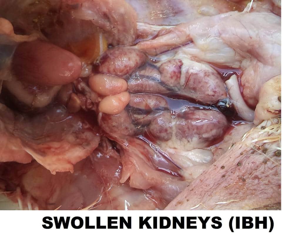
• Kidneys are enlarged, pale and mottled with multiple hemorrhages
• Lost body weight
• Diarrhea
• Anorexia
• Anemia and dehydration may develop secondary to hemorrhagic enteritis
• Sometimes the skin is icteric
• Often ecchymoses and striated haemorrhages skeletal muscles are observed.
This is particularly important in view of the fact that poultry farming has become an important livestock industry in India
Internal lesions—-
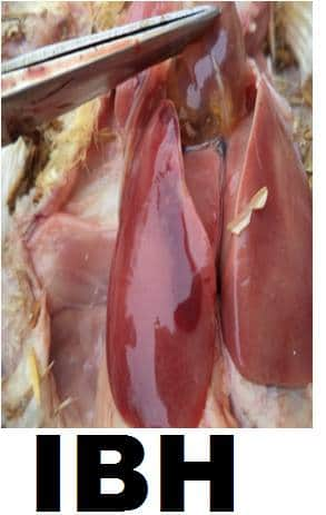
Affected chickens have mottled livers, many with pinpoint necrotic and haemorrhagic spots. Pale bone marrow and, in some cases in presence of infectious anemia, gangrenous dermatitis can be seen. Kidneys are pale and swollen. The spleen is usually quite small (atrophy). If Gumboro disease (infectious bursal disease) has been present in the birds, even if subclinical, the bursa of Fabricius will be very small (atrophic). Such chickens are immunesuppressed and usually have more severe cases of inclusion body hepatitis and/or infectious anaemia. Mature birds do not have clinical signs of adenovirus infection, they only start showing antibodies in their blood.
Post mortem lesions:—————-
• Liver swollen, yellow, mottled with petechlae and. Ecchymoses
• Kidneys and bonemarrowpale
• Blood thin
• Bursa and spleen small
Prevention & Treatment-———–
Isolate the bird from the flock and place in a safe, comfortable, warm location (your own chicken “intensive care unit”) with easy access to water and food. Limit stress.
Prevention:- ——-
• Cenrantive and good sanitary precautions, prevention of immunosuppression. Adenovirus infection may infect other organs causing a splenits inclusion body hepatitis, bronchitis, pulmonary congestion ventriculitis, oedema depending on the species of bird infected.
Reducing protein in feed in harvesting season Maize is generally contaminated we replace it with millets. Give 10 days rest to shed. we give two vaccines first vaccine company made on 2nd day & second vaccine self made (Pakistani formula) on 7th day it helps to control the disease to some extent. Avoid giving Sulpha drug in I.B.H as Anaemic like condition prevail in such disease we may add Amprosol for coccidiosis if present.
Disinfection:- ——-
Generally Iodine based disinfection like ( safe guard from vanky) or its alternative @ 1ml/3 litre of drinking water in the face of outureak & spray of 10%.formula on all the equipment curtains sheds, exhaust fans, Gunny bags etc is performed between the flocks.
Treatment:- ——–
Vaccination is only done as preventive method. now a days we got isolated cases of Hydropertcardi (Leechi disease) some home made remedies.
1) stop feed for 36 hours & give grains with salt 400 gm/qt plus Amino acids. Plus Calcium @ 500ml/1000 birds. Logic behind giving this formula is the kidney & liver are swollen & they may not digest proteins so giving grains will give some relief.
2) Give liver tonic with lipotropic factor like Choline chloride @ 200-300 ml/1000 birds along with acetic acids 15ml (500gm wt) for 4-5 days. In India homeopathic medicines are also available but their efficacy is inconsistent.
Home made (Pakistan formula) CRUDE Method:- In absence of any vaccine Dr. Qureshi from Karachi university made local vaccine which he communicated to metelephonically formula.
Liver (Affected Bird) = 80gm
+
Formalin = 2ml
+
Genetamycein 30x2ml = 60ml
+
Normal saline = 180ml
Liver will also release 30 ml of water combine all & put it mixer and grinder mix & grind for 5 minute. Then put formalin again grind. Finally put saline water & mix it for 0.5 to 1 minute. Please also see birds should not carry Gubboro or other viral disease. In such cases disease may aggravate vaccine may be injected in presence of veterinarian @ 0.2 mi/bird. After grinding 1-2 drops of formalin mix it. And again add genetamycein and mix for 1 minute then strain it with strainerused for juice straining (or Malmal cloth) twice for better result we may replace 50 ml of water with 50 ml of xlumunium Hydroxide gel for quick response. Also this vaccine may be kept at 5-6 degree celcius temperature for 12 hours before use.
vaccine prepared from one farm not be used in other farms. Potassium nitrate or Ammonium chloride can also be given at the rate of 1gm/litre.
If the disease is not complicated with complicated with coccidiosis or I.B.D it will stop with in 10 days automatically. But if the disease is complicated then in field conitions we have observed disease appearing 2-3 times in a cycle of bird.
We may also use Immunomodulators like Vitamin E. Disinfectants like VIRKON laxatives like Gur/Jaggery or any other medicine as supportive measure.
In conclusion disease can also come from as hatcheries do not inject their birds with I.B.M vaccine in slump. Bio security is the only strong tool. We may try to use branded vaccine at 1-2 days old @ 0.2ml/bird in neck. However, practically such vaccine do not work in field conditions &we may try home made vaccines. They may not harm your birds as formalin will kill the virus & gentamycein will kill all other bacteria like mycoplazma etc. Do not use old feed , do not put your old litter & avoid wet litter, do not over crowd avoid splashing of water in winter inside the shed. All such conditions promote this disease
NB-
ND + IBH KILLED VACCINE ON DAY 1 OR 3 IS ONLY PREVENTIVE MEASURES FOR IBH. It has been seen that in a flock where the vaccination has been done… the percentage of mortalies due to IBH comes down to 3 to 4 % against the 15%
Reference:On request


