Infectious Canine Hepatitis
Lalrinkima1*, R. Ralte2, S.Azmi3
1Assistant Professor, Department of Veterinary Pathology, Khalsa college of Veterinary and Animal Sciences, Amritsar
2 Associate Professor, Department of Veterinary Microbiology, Khalsa college of Veterinary and Animal Sciences, Amritsar
3 Professor & Head, Department of Veterinary Pathology Khalsa college of Veterinary and Animal Sciences, Amritsar
Infectious canine hepatitis (ICH) is a diseases of dogs caused by Canine adenovirus A (Family Adenoviridae, Genus Mastadenovirus) type 1 (CAdV-1) occurs worldwide which is more frequently observed in dogs less than 1 year of age and is clinically characterized by corneal edema. ICH has also been referred to as Rubarth’s disease, after Carl Seven Rubarth a Veterinarian who first described the disease in the late 1940s. Canine adenoviruses 1 and 2 cause severe diseases in dogs, foxes, coyotes and bears including hepatitis, ocular lesions, interstitial nephritis, encephalopathy and respiratory tract infections. In 2009, Yildirim and his co-workers reported that Canine Adenovirus (CAV) was observed for the first time in Kars Shepherd Dogs. ICH virus has been associated with other disease syndromes in dogs including encephalopathy, iridocyclitis, interstitial nephritis and with respiratory disease.
Clinical signs
Clinical symptoms of infectious canine hepatitis include fever, inappetance, diffuse hemorrhages, abdominal pain, vomiting, diarrhea and less frequently dyspnea. Corneal opacity (‘‘blue eye’’) and interstitial nephritis may occur 1–3 weeks after the clinical recovery as a consequence of the deposition of circulating immune complexes. Rarely, neurologic signs such as seizures, ataxia, circling, apparent blindness, head pressing and nystagmus have been reported in association with CAV-1 encephalitis.

Figure 1: Corneal edema
Postmortem findings
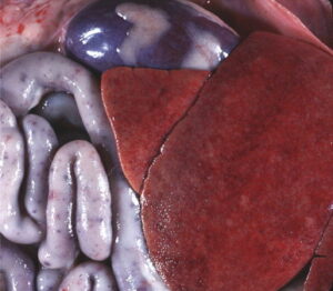
Gross pathologic findings in dogs with ICH include blood-tinged ascites or hemoabdomen, a slightly enlarged, congested or mottled liver, mild splenomegaly, enlarged, congested and edematous lymph nodes and fibrin deposition on the surface of abdominal viscera. The gallbladder wall is typically markedly thickened and edematous. Petechial and ecchymotic subserosal hemorrhages may be apparent in multiple viscera and the intestinal tract may contain bloody fluid. The brain may also contain petechial hemorrhages or areas of gray discoloration.

Figure 2: The liver from dogs with ICH can be slightly enlarged and friable with a blotchy yellow discoloration
Diagnosis
CAdV-1 infection was confirmed by quantitative real-time PCR of cell suspension. Postmortem findings and histopathological changes are highly suggestive of CAV-1 infection indicated that there was presence of intranuclear inclusions in liver. Confirmation of a diagnosis of ICH is obtained by virus isolation on permissive cell lines such as Madin Darby canine kidney (MDCK) cells. A polymerase chain reaction (PCR) protocol has been used for molecular diagnosis. Ocular swabs, feces, blood, tissues and urine samples can be collected in vivo for virus isolation and PCR.
References
- Caudell D., Confer A.W. and Fulton R.W. (2005). Diagnosis of infectious canine hepatitis virus (CAV-1) infection in puppies with encephalopathy. J Vet Diagn Invest. 17: 58–61.
- Decaro, N., Campolo, M., Elia, G., Buonavoglia, D., Colaianni, M. L., Lorusso, A., Mari, V. and Buonavoglia, C. (2007). Infectious canine hepatitis: an ‘‘old’’ disease reemerging in Italy. Res. Vet. Sci., 83: 269–273.
- Rubarth, S. (1947). An acute virus disease with liver lesions in dogs (Hepatitis contagiosa canis). A pathologico-anatomical and aetiological investigation. Acta Pathol. Microbiol. Scand., 69: 1.
- Yildirim, Y., Kirmizigul, A. H. and Gökçe, E. (2009). Seroprevalence of Canine Adenovirus (CAV) Infection in Kars Dogs in Turkey. Y. Y. U. Vet. Fak. Derg. 20: 37 – 39.

