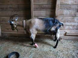
LAMENESS IN GOAT & SHEEP : MANAGEMENT & TREATMENT
Abnormality of gait is a sign common to many diseases and conditions. A complete history is important for diagnosis and should include incidence and duration in the herd, nutrition, feed changes, method of rearing, and recent introductions to the herd . Some causes of lameness may be associated with systemic disease. Therefore, a thorough physical examination should always be performed, followed by a detailed examination of all four limbs, with a specific assessment of gait and mobility in an attempt to localize locomotor problems. In goats, as in other species, locomotor difficulties usually involve the musculoskeletal system directly, but conditions of the nervous system can mimic musculoskeletal disease and should be considered during the clinical examination. Some of the more important conditions that cause lameness in goats are discussed below—
Signs associated with lameness: (lower & upper leg)———
Stiff, painful gait with shortened stride such as foot rot/scald; arthritis Non-weight bearing; single leg for example fracture Paresis; stumbling, reluctance to rise such as caused by anemia Walking on knees including CAE Tendon and joint capsule contracture
Causes of lameness ———
Heritable Chondrodysplasia/ Spider Lamb syndrome —-
Mainly in Suffolk and Hampshire breeds Mutation in fibroblast growth factor receptor (DNA test available) Autosomal recessive (two copies of an abnormal gene must be present) Signs may be present at birth or may develop later (by week 6) Chondrodysplasia is defined as exostoses at the epiphyses resulting in arrested development and deformity Angular limb deformities are common
Myotonia Congenita /Fainting goats———–
Heritable disorder (autosomal dominant trait) Mutation in the chloride channel in skeletal muscle Hyper excitability of the sarcolemma resulting in delayed relaxation Tetanic muscle contraction when startled (may collapse)
Luxation of the Patella —————
Can be congenital or genetic predisposition Unable to hold the stifle in extension Diagnosis: Patella easily dislocates medially or laterally Treatment: Joint capsule imbrication lateral/release medial/TPLO/Trochlea implant
Carpal Contracture ————–
Can be congenital in goats Secondary to injury/disease (CAE) that prevents weight bearing Fibrosis of joint capsule Treatment: Mild: splints and bandages Tenotomy of flexors Poor prognosis with joint capsule fibrosis
Predator Attack———–
Mainly dogs and coyotes Animal in shock. High mortality Damage to deeper structures hard to asses Treatment: IV Fluids/ steroids/ Aggressive pain and anti-inflammatory treatment Tetanus toxoid/antitoxin Antibiotics Clean/debride wounds Pain control Prolonged recovery/high cost
Claw overgrowth due to inadequate care/laminitis————-
Distortion of the claw capsule 307 adult goats from 4 large commercial dairy farms was examined. Horn overgrown 83 – 95% in at least one foot. Trimming Steps Clean claws and interdigital skin Look for swelling, loose horn, lesions 1st cut: tip of claw to sole depth 2nd cut: abaxial wall level with sole 3rd cut: axial level with sole
Corrective trimming————–
Remove all loose and undermined horn.————
If exposed corium, anesthesia of the leg is required as follows: Apply tourniquet to just below elbow or hock. Inject 1-3ml lidocaine IV below tourniquet Combine with 0.1mg/kg Xylazine IM or 1-2ml diazepam IV or Ket/diazepam IM Apply orthopedic foot block if indicated to relieve weight bearing
Degenerative joint disease—————-
Chondroprotective agents Polysulfated glycosaminoglycan (Adequan) 125mg/week for 5 weeks IM Hyaluronic acid Normal joint lubricant/increases viscosity Inhibit bacterial growth Methyl Sulfonyl Methane MSM Source of Sulphur Oral and/or Intra-articular steroids Supportive treatment Physical therapy Floating Sling Massage/ movement Electro-acupuncture
Laminitis —————–
Etiology/Pathophysiology
Laminitis in small ruminants can be caused by infectious, toxic or nutritional and management factors. Infectious causes include bluetongue and foot and mouth disease. Blue tongue causes damage to vascular endothelium, which in the feet results in coronitis. Foot and mouth disease can cause interruption in horn growth resulting in lameness and the development of a horizontal groove in the wall. This is seen in antelope and African buffalo in foot and mouth endemic areas. Acute laminitis can result from over consumption of grain, however subclinical or chronic laminitis with the development of secondary claw horn lesions is more common. This includes abnormal growth with distortion of the hoof wall, horizontal or vertical wall cracks and fissures, generally poor quality horn and white line disease. Inadequate claw care may result in overgrowth and loss of normal claw conformation with lameness as a result. Toxic causes of laminitis may include severe systemic disease such as mastitis or metritis or metabolic cause such as pregnancy toxemia. Plant toxins include alkaloids from different Crotolaria species. The most common is Crotolaria burkeana which causes laminitis in small ruminants in sheep and goats and cattle in Southern Africa and India
Systems Affected ————-
Depending on cause———
Locomotor: Inflammatory changes in different parts of the corium resulting in abnormal proliferation, differentiation and keratinization of the epidermis
Signalment / History———
Will depend on geographical location, nutrition and management and presence of infectious agents
Clinical Features————
Lameness in sheep and goats can result in significant production loss since less time is spent eating and they have a reduced ability to compete for food with their healthy flock mates. Lame sheep and goats spend more time lying down or grazing while on their knees. They rapidly lose body condition. Poor body condition leads to reduced sperm production and therefore decreased fertility. Similarly lameness also leads to lower ovulation rates and lower birthing percentages In acute laminitis the coronary band may be red and swollen and the feet warm to the touch. The animal may shift weight around when standing and show lameness in one or more feet during movement. In subclinical cases the feet show signs of claw horn disruption such as white line separation and wall cracks.
Differential Diagnosis ————–
White line degeneration with necrotic tracts extending between the wall and sole White line and subsolar abscess with extensive white line separation between the wall and corium (shelly foot/claw) Sepsis of the distal interphalangeal joint; Gross claw overgrowth leading to wall horn fracture and exposure of the corium Upper leg problems including injury and joint-ill
Diagnosis————–
Diagnosis is based on physical examination and detailed history. Radiographs are necessary if the foot is swollen. In cases where the source of lameness cannot be determined local regional anesthesia below a tourniquet is useful to rule the lower limb out as the source of pain.
Treatment —————-
The cause of the lameness should be identified and removed. Corrective trimming to remove loose horn such as caused by white line disease should be carried out. All loose horn is removed until reattachment with healthy horn. Overgrowth with restoration of normal weight bearing surface and claw conformation should be carried out if possible. However, over aggressive trimming can induce severe lameness and delay healing. Application of a modified foot block can be used to remove weight bearing from an affected claw (S Van Amstel – unpublished clinical cases). Use of antibiotics should be restricted to cases where secondary infection is present. Long-acting antibiotics such as oxytetracycline or tulathromycin are commonly used. Daily injections of procaine penicillin have also shown to be effective against bacterial infections of the foot. Amputation of the digit can be performed where infection has penetrated the distal phalanges or joints of the foot. Palliative treatment to control pain is generally used. Non-steroidal anti-inflammatories such as flunixin 1.1 mg/kg given IV once or twice daily or meloxicam 0.5 mg/kg orally once daily are commonly used.
Interdigital dermatitis (ID)-————-
Key facts: F necrophorum & A.pyogenes. Excessive moisture and fecal contamination. F necrophorum can survive months in fecal contaminated, muddy environment. Clinical signs: Lameness – variable; Interdigital skin red, swollen, erosions; Pitting of soft horn.
Control: Environment should be kept clean and dry. Foot baths; weekly; 10% Zinc sulfate or 5% Formaldehyde. Systemic Penicillin in severely affected animals
Foot abscess—————
Key facts.
F. necrophorum & A. pyogenes. Acute or chronic, purulent non-contagious disease. Follows ID or interdigital trauma. Involves one digit – DIP joint. Low prevalence. Epidemiology: Interdigital skin maceration and trauma. Seasonal association with tick bites. Pathogenesis: Infection enters through skin and joint pouch-Septic arthritis. Eventual fibrosis and ankylosis. Deformity of affected digit Diagnosis: Swelling above coronary band; Sinus tracts at coronet or interdigital skin; Discharge of creamy-white pus; Claw usually normal; Treatment: Antibiotics; Trephine, curettage, lavage; Remove weight-bearing. Application of claw block to healthy claw; Claw amputation
Foot rot; ——————-
Key facts.
Dichelobacter nodosus & F.necrophorum Highly contagious painful & debilitating necrotizing inflammation of the interdigital skin and horn with necrotic odor; Different strains of D. nodosus. Wet, muddy conditions/interdigital trauma/ ambient temp above 10oC. 3 forms: Benign (non-progressive) BFR; Intermediate IFR, Virulent (progressive) VFR.
Epidemiology.
Asymptomatic carrier sheep main reservoir Cattle can be source. D. nodosus obligate parasite of skin of feet but does not survive >7-14 days away from host. Carriers remain infected for 2-3 years
Diagnosis:
Scoring system: 0 = normal; 1 = mild localized ID ; slight/moderate inflammation and erosion; 2 = necrotizing inflammation extends to part or all of the soft horn of the axial wall; 3 = involvement of the heel and sole horn ; 4 = necrotizing inflammation extending to the laminae under both walls. Objective of flock diagnosis/ scoring system: Economically important vs. trivial impact.
Trimming:
Remove all loose and under-run horn. Foot bathing once a week during high risk period. Zinc/Copper/Formaldehyde. Contact of foot bath solution for 5 minutes. Keep in dry area 10-20 minutes.
Antibiotic treatment:
Procaine Pen 20-30,000u/kg bid; Erythromycin 3-5mg/kg bid; Zactran; Oxytet LA 20mg/kg every 48 hours; Tulatromycin. Severely affected animals should be culled.
Eradication:
Method 1. Flock disposal/ replacement with clean animals after 2 weeks Method 2. Carried out when transmission is unlikely to occur. Inspect all feet of all animals 3 times at 3 weekly intervals and look for VFR/IFR. Dispose of infected animals. Method 3. Identify and treat all affected animals. Inspect all feet of all animals 3 times at 3 weekly intervals and treat. Inspect treated animals after 3 weeks. Cull non-responsive cases.
Post eradication: Continual monitoring/re-examination of animals considered free of FR. Eradication successful when flock remains free after at least 1 transmission period
Septic arthritis.—————–
C pseudotuberculosis (CLA); Haemophilus; M haemolytica; T pyogenes; Erysipelothrix rhusiopathiae; Chlamidia in sheep feedlots. Umbilical infection/tail docking.
Mycoplasma associated with septicemia, pneumonia, poly-arthritis, mastitis. Transmitted through infected milk; mites; fleas. Kids 3-8 weeks most susceptible. Mycoplasma occur in 3 forms 1. Peracute septicemia. Death in 12-24 hours. 2. Brain form. Neuroligical signs (opisthotonus). Death in 24-72 hours. 3. Septic arthritis and pneumonia.
Treatment.
Antibiotics does not eliminate infection. Tylosin 10-50mg/kgTID; Oxytetracycline; Florfenicol; Zactran; Tulathromycin
Viral causes of lameness: ————–
Blue tongue; Foot and mouth disease; Ulcerative dermatosis; Vesicular stomatitis; CAE (Caprine Arthritis Encephalitis).
CAE: ——————–
Retro virus; Clinical disease; heavily infected flocks 10%.
Epidemiology:
Transmission mainly through colostrum. Carried in cells in milk but also present in cell free milk. Can be transmitted by a single feeding. Risk of infection low if removed immediately after birth. Intrauterine transmission possible. Also peri-natal contact with secretions. Horizontal transmission can occur at all ages with prolonged co-mingling/Introduction of susceptible animals. Antigenic drift common; facilitate persistence in host in monocytes and macrophages. Neutralizing antibodies has no effect on the condition.
Pathogenesis:
4 syndromes:
Chronic arthritis; Indurative mastitis; Chronic interstitial pneumonia; Encephalitis.
Clinical Findings:
Arthritis involving mostly the carpal joints/ uni- or bilateral with intermittent lameness and weight loss. Radiographs show soft tissue swelling and calcification of peri-articular tissues with osteophytes in chronic cases. Synovial fluid increased mononuclear cell count. Kids 1-5 months can develop a leukoencephalitis and show neuro signs such as posterior paresis and ataxia; head tilt; torticollis; circling; tetra-paresis; may remain bright and alert and eating. Indurative mastitis; Firm and hard udder; no milk. No systemic illness
Diagnosis:
Cerebrospinal fluid:
Increase mononuclear cells. Serological testing: AGID – Positive test older than 6 months = infection. Majority sero-positive for life. Virus isolation.
Differential Diagnosis:
Copper deficiency (Sway back). Spinal abscess; Meningeal worm; Listeriosis; Polioencephalomalacia; Infectious arthritides; Mastitis
Control:
Remove kids at birth; prevent contact. Heat-treated goat colostrum then replacer All animals over 3 months test 6 monthly; cull or segregate all sero-positives. Herd accreditation. 2 negative tests. Restriction on purchase and movement
Nutritional causes of lameness.——————–
Osteodystrophy; Ca:P04 imbalance; Copper deficiency; Hypovitaminosis D, Lead; Fluorine toxicity.
Angular limb deformity:
Heritable; rapid growth
Contagious ovine digital dermatitis—————-
Caused by Spirochetes Other causes of Lameness Interdigital “balling” of grass and dirt Strawberry foot rot (combined orf and Dermatophilus infection).
Post-dipping lameness – cellulitis and polyarthritis due to Erysipelas rhusopathiae from contaminated dips
Septic Arthritis—————–
Bacterial
C pseudotuberculosis (CLA) Haemophilus M haemolytica T pyogenes Erysipelothrix rhusiopathiae Umbilical infection/tail docking Chlamidia Sheep feedlots Mycoplasma Infected milk; mites; fleas Kids 3-8 weeks most susceptible Septic arthritis 3 Forms Peracute septicemia. Death in 12-24 hours Brain form. Neuroligical signs (opisthotonus). Death in 24-72 hours Septic arthritis and pneumonia
Treatment:
Mycoplasma Antibiotics does not eliminate infection Tylosin 10-50mg/kgTID Oxytetracycline Florfenicol
White Muscle Disease ——————
Nutritional Muscular Dystrophy Deficiency in selenium and vitamin E Most common in young, rapidly growing animals Ill thrift, reproductive losses Faster growing plants have less selenium Other minerals or feed contaminants can hinder selenium absorption Forage with less than 0.1 ppm (on dry matter) is considered deficient Colostrum is rich in vitamin E Acute (cardiac) form: recumbency, respiratory distress, death Subacute (skeletal) form: stiff gait, tremble while standing
Diagnosis:
elevated CK, AST Blood selenium (>95% of selenium is inside the RBC) Serum tocopherol Necropsy: friable muscles, pale streaks
Treatment:
Vitamin E/Selenium injection + oral vitamin E Most respond to treatment
Prevention:
Supplement diet with selenium/Vit. E (especially in pregnant animals) Monthly Vit. E/Selenium injections
Compiled  & Shared by- Team, LITD (Livestock Institute of Training & Development)


Image-Courtesy-Google


Reference-On Request.


