Malassezia Dermatitis (Yeast Infections) in Dogs: Causes, Treatments, Prevention
COMPILED & EDITED BY-DR. ASHUTOSH MISHRA,PATNA
Malassezia dermatitis is a very common skin condition also known as yeast dermatitis. It’s caused by the Malassezia pachydermatis fungus, which is normal to have on the skin. Problems only arise when it grows excessively and leads to inflammation of the skin, more commonly known as dermatitis. This condition can be quite itchy for your dog and it requires long-term treatment, though most dogs will recover just fine and the itchiness should abate in the first week of treatment.
Malassezia pachydermatitis is the name of a species of yeast that normally lives on the surface of the canine skin. In the presence of allergic skin disease or any other kind of inflammatory skin disorder, this microorganism has a tendency to proliferate abnormally. Waxy, oily, scaly and/or moist skin––including the skin that lines the ear canal––is particularly predisposed. Malassezia dermatitis (MD) is a superficial fungal (yeast) infection occurring on and within the stratum corneum of the epidermis of many mammalian species. There are several different species of Malassezia yeast recognized, and various animals may serve as the natural hosts for specific species of the yeast. For example, most domestic carnivores harbor Malassezia pachydermatis as part of their natural cutaneous microflora, while human beings primarily harbor Malassezia furfur. As commensal organisms, Malassezia yeast colonize the skin in very low numbers. Overt infection is defined by increased numbers of the yeast on the skin surface in conjunction with inflammation. In dogs with atopic dermatitis (AD), M. pachydermatis may be recognized by the immune system as an allergen, in which case a highly inflammatory and pruritic response can be mounted to relatively low numbers of yeast organisms, blurring the line between “colonization” and “infection.” The role of M. pachydermatis in feline dermatitis is less well defined, although it is a known commensal of feline skin as well. Yeast dermatitis or Malassezia dermatitis as it’s scientifically known is a fungal infection of the skin that occurs when a dog’s immune system becomes compromised. It can range in severity from mild to extreme and seems to affect certain breeds more than others. Treatment can take the form of oral medication or anti-fungal shampoos, though dogs with severe yeast dermatitis will likely require both. It’s a very treatable condition, but if left untreated it can severely impact your dog’s quality of living.
The genus Malassezia includes 6 species of lipodependent yeasts and Malassezia pachydermatis which is lipophilic but not lipodependent. The reproduction of Malassezia is asexual with a unipolar budding. This gives a typical shape (resembling a footprint or a peanut…). Their size is small (2 to 7 µm). They do not form pseudomycelium. Round, convex and yellowish cultures develop on Sabouraud’s dextrose agar.
Many studies have shown that Malassezia pachydermatis is a component of the normal cutaneous flora of the dog, using various techniques. Around 50 % of healthy dogs are carriers of this yeast which can be found in the external ear canal, the skin (particularly the anal area which could be a carriage zone, the lips and extremities) and the haircoat. Malassezia could survive in the gastrointestinal tract since it can be isolated in the faeces. Only a few authors have isolated lipodependent yeasts (M. furfur, M. sympodialis) without indicating the technique that was used or with a peculiar technique.
The response of the host to the yeast includes non-specific defense mechanisms (phagocytosis by neutrophils) as well as cell-mediated specific defense mechanisms. In the latter, Langerhans cells present the antigen which activates T-cells. These T-cells multiply and produce lymphokines which stimulate phagocytosis by macrophages and multiplication of epidermal basal cells. This leads to the destruction of the yeasts or to their mechanical removal with scaling.
Malassezia pachydermatis infection has been recognised as a major cause of skin problems in canine practice. The infections can be primary, or secondary to underlying allergic, endocrine or neoplastic conditions. The infection is a major cause of pruritus in dogs and yet it is often overlooked because there is a tendency to concentrate on the allergic diseases which often are also present. Malassezia spp are unicellular budding yeast organisms that divide asexually and are classified as lipid-dependent or nonlipid dependent organisms, based on their growth requirements. Several different Malassezia species have been isolated from skin, but of these, Malassezia pachydermatis, a non-lipid dependant species, is the most studied species in veterinary medicine. M. pachydermatis is considered to be commensal on canine and feline skin, but can cause infections when the microclimate on the skin surface, or in the ear, is altered, or if the host immune responses are compromised. Other, lipid-dependent species, M. sympodialis, M. globosa, M. furfur, and M. sloofiae have been isolated from cats and dogs.
Alterations of the cutaneous microclimate or host defense mechanisms allow Malassezia pachydermatis to multiply and to become pathogenic. Cutaneous factors enhancing the multiplication of Malassezia pachydermatis are: an excessive production or a modification of sebum and/or cerumen, an excess of moisture, a rupture of the epidermal barrier and cutaneous folds.
These changes may be due to underlying causes, of which the following are most common:
Cutaneous hypersensitivity including atopic dermatitis,
Pyoderma,
Ectoparasitic skin disease, particularly demodicosis,
Endocrine disorders, particularly hypothyroidism,
Keratinization disorders: epidermal dysplasia of the West Highland white terrier, idiopathic seborrhoea,
Treatment with glucocorticoids or antibiotics.
Immunological dysfunction (cell-mediated immunity, IgA secretion) could also promote the growth of the Malassezia population on the skin and its pathogenicity. For instance epidermal dysplasia of the West Highland white terrier could be associated with a genetic predisposition to a poor response of T-cells towards the yeast. Nevertheless the epidermal hyperplasia seen in Malassezia dermatitis is not caused by secretion of products from the yeast.Malassezia produce many enzymes (including lipases and proteases) which can contribute to cutaneous inflammation through proteolysis, lipolysis (which alters the lipidic cutaneous film), changes of cutaneous pH, eicosanoid release and complement activation.
In addition, it has been shown that Malassezia pachydermatis could play an allergenic role. There is a type 1 (immediate) hypersensitivity. Skin-testing with a Malassezia extract can show immediate hypersensitivity reactions. In addition, levels of specific IgG and IgA are greater in dogs with Malassezia dermatitis than in normal dogs. There are higher levels of specific IgG and IgE in atopic dogs (with or without concurrent Malassezia dermatitis) than in non-atopic dogs with Malassezia dermatitis/otitis and normal dogs. Recently, the functionality of anti-Malassezia IgE has been demonstrated through passive transfer using the Prausnitz-Küstner technique. Some major allergens of Malassezia pachydermatis have been identified: proteins with 45, 52, 56 and 63 kDa molecular weight. The delayed hypersensitivity is less known: the in vitro proliferative response of peripheral blood mononuclear cells in dogs with Malassezia dermatitis does not exceed the one of healthy dogs. Delayed reactions (24 hours) to skin tests exist in basset hounds with or without Malassezia dermatitis and are more frequent than in healthy beagle dogs.
Epidemiology
There is no age or sex predilection except in one study that demonstrated a predisposition of neutered male and female dogs. Some breeds are predisposed to Malassezia dermatitis: West Highland white terrier, Basset hound, English Setters, Shih Tzus, Dachshund, Cocker spaniel, American Cocker Spaniel, Poodle, German shepherd, Collies, Shetland, Jack Russell terrier, Silky terrier, Australian terrier, Springer spaniel and Shar-Pei.
Malassezia dermatitis is often seasonal (from the end of spring to the beginning of fall which is the time at which allergic dermatites are often diagnosed). It can persist during the winter.
There is no indication that Malassezia dermatitis is contagious.
Clinical signs
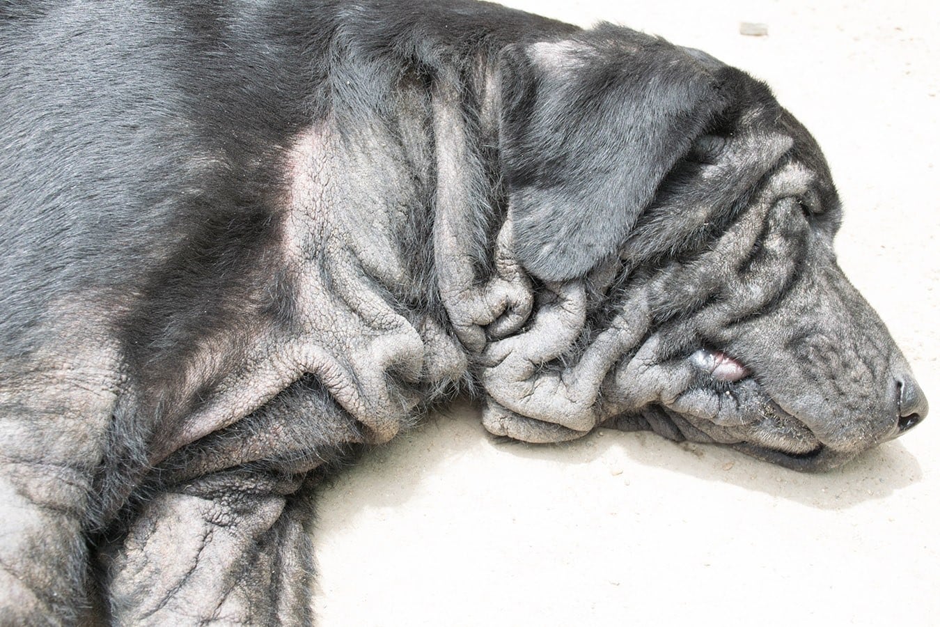
Pruritus is always present and severe. Animals are presented with a strong odour of rancid fat.At the beginning of the disease there are localized or diffused erythema, erythematous papules and macules, and a keratoseborrhoeic disorder with scaling, crusting and alopecia and a greasy aspect of skin and hair.This is followed rapidly by secondary lesions such as lichenification and hyperpigmentation.Malassezia dermatitis can be localized, e.g., on the ventral side of the body (neck, axillae, ventrum and inguinal area), face (ear pinnae, lips, muzzle), peri-anal area and legs (forearms, caudal thighs and feet). It can also be generalized. It is not uncommon to observe concurrent otitis externa.Lymph node enlargement is sometimes seen but most often there are no general signs.
The most common signs of this condition include:
- Itchiness
- Red skin
- The dog is emitting a musty odor
- Increased dark pigment in the skin
- Chronic ear infections
- The skin becomes thick
- Crusty, flaky skin with scales
Certain canine breeds seem to experience higher rates of yeast dermatitis than others.
Breeds that are considered to be the most at-risk for this condition include:
- Dachshunds
- Australian Terriers
- Basset Hounds
- West Highland White Terrier
- Chihuahuas
- Cocker Spaniels
- Shih Tzus
- English Setters
- Silky Terriers
- Shetland Sheepdogs
- Boxers
- Lhasa Apso
- Maltese Terriers
- Poodles
Diagnosis
Diagnosis of Malassezia dermatitis is based upon history, physical examination, appropriate complementary diagnostic aids to show the presence of Malassezia on the skin, response to specific therapy and exclusion of other dermatoses.
Cytological examination
Cytological examination can show yeasts and allow for a semi-quantification. The result is to immediately us the immersion power objective after staining with lactic blue or, preferably, a rapid staining method. Several cytological techniques can be used:
Impression smear
“Scotch test” using pieces of tape (clear cellophane) strip
Scrape smear
Swab smear
In many hands, impression and above all tape strip smears appeared to be the most reliable methods. Swab smears should be reserved for cytological examination of the external ear canal. Cytological examination will show oval or elongated cells of 3 to 5 µm in diameter, with a typical single polar budding (“footprints, peanuts, Perrier bottles”…). Yeasts can adhere to scales. A suppurative reaction is not uncommon.
The minimal number of yeasts which indicates the possibility of a true Malassezia dermatitis is not really known. Some authors feel that even a few yeasts are significant whereas others would consider the disease only if there is a higher number of yeasts per high power field. Perhaps the number of yeasts is an indication. In addition there are variations between breeds and body sites. Lastly there are cases in which a small number of yeasts triggers a hypersensitivity reaction and so the ultimate criterion will be the response to antifungal therapy.
Fungal cultures
Fungal cultures can show the presence of Malassezia on the skin and hair of dogs. Sampling can be done with hairs, swabs, pieces of rugs, contact plates or liquid detergents. Appropriate media for Malassezia pachydermatis are Sabouraud’s dextrose agar with chloramphenicol and cycloheximidine (which improves the growth of the yeast) and modified Dixon’s agar which grow all species of Malassezia. As the yeast is a normal component of the cutaneous flora of the dog, by itself a positive culturing has no or little value. However, the number of colonies is perhaps an indication, as for all opportunistic agents (this is comparable to the number of yeasts demonstrated by cytological examination).
Cutaneous histopathology
Cutaneous histopathology can sometimes show the yeasts on the surface of the epidermis and in the infundibula, particularly in PAS stained sections (although they are occasionally visible on HE stained sections). However if they are not discovered this does not exclude their presence (biopsy in a non-infected area, removal of the stratum corneum during processing, etc). Cutaneous histopathology is a less sensitive technique than cytology. As for cytology the presence of the yeast on the skin may have a variable meaning since it can be discovered in normal dogs and dogs with various dermatoses. In contrast, the finding of Malassezia inside hair follicles could indicate a real pathogenicity.
There are common findings in biopsies from dogs with Malassezia dermatitis, leading to a pattern including:
Orthokeratotic hyperkeratosis with prominent foyers of parakeratosis,
Acanthosis and spongiosis with irregular rete ridges,
Lymphocytic exocytosis of the epidermis,
Intraepidermal neutrophilic or eosinophilic pustules,
Moderate dermal inflammatory reaction, perivascular to diffuse, with lymphocytes, plasma cells, histiocytes and often neutrophils, eosinophils and mast cells,
Subepidermal linear alignment of mast cells (SLAM).
Signs of concurrent bacterial folliculitis are not uncommon. Rarely, folliculitis and furunculosis can be observed in association with the presence of yeasts inside the air follicles.
Therapeutic challenge
The therapeutic challenge is in fact the ultimate tool to confirm that in a particular case the commensal Malassezia has become a pathogen, thereby playing a role in the development of the dermatitis.
Differential diagnosis
Differential diagnosis includes many pruritic dermatoses with erythema, hyperpigmentation and seborrhoea including allergic skin diseases, bacterial folliculitis, demodicosis, scabies, drug reaction, idiopathic acanthosis nigricans, epitheliotropic lymphoma and all causes of seborrhoea with cutaneous inflammation. In fact clinical signs of Malassezia dermatitis are so variable that it may mimic many dermatoses. Furthermore, Malassezia dermatitis is often associated to or even promoted by most of the dermatoses which are included in its differential diagnosis.
Treatment
Systemic therapy
Systemic therapy is necessary in many cases, particularly when clinical signs are severe and when the lesions are extensive. Ketoconazole is the most commonly used drug. As with all azole derivatives, ketoconazole acts in binding to cytochrome P450, which inhibits synthesis of ergosterol, an important component of the fungal cell membrane. This results in alterations of cellular permeability and activity of various membrane enzymes. Ketoconazole also has anti-inflammatory properties through an action on leucotriene synthesis and it has an action on the keratinization process through an action on alltrans retinoic acid. The dose is 10 mg/kg/day (the author would not however give more than 200 mg/day, the human daily dose, to a dog). It is recommended to give the drug with some food. Tolerance is usually good but periodic biochemistry panels are necessary during long treatments. In effect, an increase in serum transaminases may be followed by signs of intolerance (anorexia, vomiting) due to hepatic toxicity. Itraconazole could also be used (5 to 20 mg/kg every day or other day). To our knowledge, Malasseziapachydermatis has not shown any resistance to antifungal agents commonly used against yeasts (azole derivatives, nystatin, amphotericin B, 5- fluorocytosine). Griseofulvin and allylamine derivatives are not effective in treating Malassezia.
Topical therapy
Topical therapy is an alternative to systemic treatment, particularly for localized lesions (creams, gels, lotions or sprays). For extensive lesions antifungal shampoos or lotions are preferable. They can be used with systemic therapy, although there is no formal evidence that the combination is of greater value than systemic treatment alone.
Topical therapy alone should not be used as a diagnostic challenge, but it can maintain a remission, thus confirming the diagnosis. Shampoos containing miconazole (2%), chlorhexidine (at least 3%), a combination of both (2% each) and ketoconazole (2%) are the best whereas the most appropriate leave-on rinses (lotions) are lime sulfur and above all enilconazole (10 % diluted 50 times i.e., 0.2 %). Topical treatments should be administered 2 to 3 times a week during 2 weeks then once a week.
Therapeutic follow-up
Therapeutic follow-up is very important. First of all, an improvement confirms the diagnosis. Pruritus usually decreases within one week, whereas lesions will clearly decrease after 2 weeks of treatment, particularly if both systemic and topical therapies are used. The duration of treatment should be at least one month and can be as long as 2 months to get a complete recovery. Usually therapy is continued for 7 to 10 days beyond clinical cure. Otitis externa should be treated vigorously to limit the fungal reservoir (nystatin, thiabendazole, clotrimazole, miconazole, antiseptic cleansing agents, etc). In cases of concurrent superficial pyoderma or bacterial overgrowth (BOG), antibiotic therapy should be used simultaneously. Malassezia dermatitis being often secondary to an underlying dermatosis, it is important to diagnose and treat it appropriately. In case of “idiopathic” Malassezia dermatitis or if such a control is impossible, relapses can be prevented either by weekly topical treatments or by oral administration of ketoconazole 1 or 2 days a week.
References
References are available upon request.
https://www.pashudhanpraharee.com/major-skin-problems-in-dog-causessymptomsdiagnosis-treatment/
https://vcahospitals.com/know-your-pet/yeast-dermatitis-in-dogs


