MAMMARY TUMOURS IN DOGS AND CATS
Mammary tumours are extremely common in dogs and cats, and these can be emotive cases as clients are more aware of breast cancer and may have some personal experience.
Throughout the oestrus cycle there is significant development and regression of the mammary tissue that can be observed histologically in dogs and cats. For this reason, normal mammary tissue can look very different histologically depending on the stage of oestrus or neutering state.
There is a lot of data regarding risk factors for developing tumours in women and the same processes have been applied in animals. These include: increasing age, with an increase in tumour formation beyond about seven years in the dog and nine years in the cat; age at neutering; breed susceptibility/genetics; exogenous hormone therapy; and diet/obesity. For dogs and cats, pregnancy, parity and pseudopregnancy in the bitch do not appear to affect the risk of developing mammary tumours.
There are classification systems for naming mammary tumours in both dogs and cats. Over 90 percent of mammary tumours in elderly cats are malignant. Canine mammary tumours are more diverse with more benign tumours recognised, including the very common mixed and complex adenomas. Roughly 45 percent of these are malignant.
Breast cancer in women is graded using the Elston-Ellis (or Nottingham) grading system. We can grade both canine and feline mammary tumours using modifications of this system. Tumours are graded based on tubule formation, degree of nuclear pleomorphism and mitotic count per 10 high-power fields to give a total score. From the total score tumours can be graded as 1: low-, 2: intermediate- and 3: high-grade.
Other prognostic indicators include tumour diameter. Tumours greater than 3cm diameter in dogs and 2cm in cats tend to have a poorer outlook. Further clinical staging includes evidence of lymphovascular invasion and lymph node spread. Obviously further metastatic spread to the lungs is indicative of a poor prognosis.
Immunohistochemistry has been used to subdivide breast cancers in women and has prognostic value. Common markers include oestrogen and progesterone receptors and human epidermal growth factor receptor 2 (HER2). Recently there have been attempts to classify canine and feline mammary carcinomas by similar molecular subtypes. The outcome has varied depending on the study, probably due to the different panels of antibodies used and, although the subtypes were identified in some studies, the results are contradictory and additional investigations are being done to try to standardise protocols. For the time being, in dogs we use COX-2 and in cats COX-2 and Ki67 as prognostic markers.
Surgery remains the mainstay of treatment, with oncologists also using chemotherapy and/or radiotherapy as adjuvant therapy and for palliative care to reduce pain and shrink tumours.
SIGNS
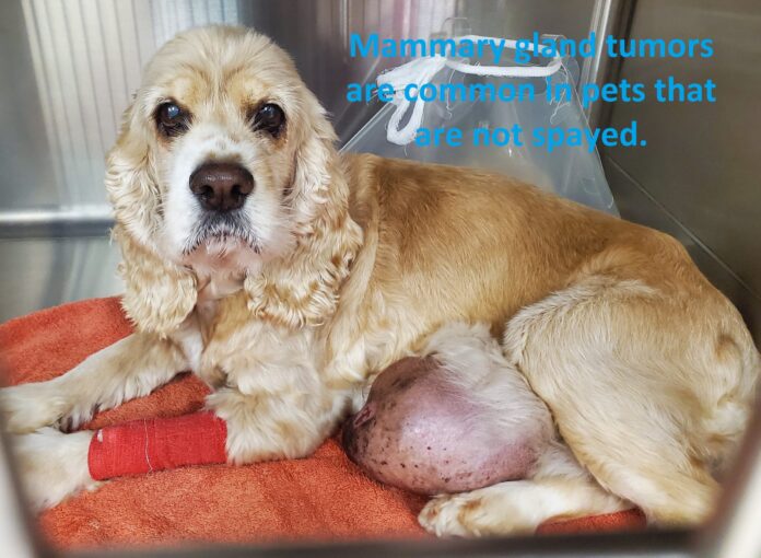
Mammary tumours are usually first detected as small, nobbly lumps underneath the skin, located in and around the mammary glands. Sometimes the tumour will ulcerate and become painful and inflamed. The rate of growth and spread varies with the type of tumour.
The extensive tumours pictured here would have been growing for some time. Leaving lumps to grow and not seeking veterinary advice when first noticed – regardless of whether they are benign or not – makes surgery much more challenging and problematic, both for the surgeon and the pet. Recovery takes much longer and the pet will need more intensive post-surgery care and pain relief.
Classification of Mammary Tumors
Mammary tumors can be malignant or benign. In dogs, up to 50% are malignant. In cats, almost all mammary tumors are malignant (adenocarcinomas). Although there are histologic variations on this, these are the main classifications. The more common ones are at the top of each list:
Benign
- Adenomas
- Mixed tumors
- Fibroadenomas
- Mesenchymal
Malignant
- Tubular adenocarcinomas
- Papillary adenocarcinoma
- Anaplasric carcinoma
- Sarcomas
- Solid carcinomas
- Mixed
Causes: Mammary tumors develop because of spikes in female hormone (estrogens) that take place during a dog’s heat cycle. By spaying a dog at 6 months of age or before the first heat cycle, it virtually eliminates the risk of getting mammary tumors, which starts at only about 0.5%. Once a dog goes through one single heat cycle, the risk increases to 8%. After a second heat cycle, the risk shoots up to 26% (says the American College of Veterinary Surgeons).
If a dog is spayed after 2 years of age, then there is no more protection. Over 25% of non-spayed female dogs will develop mammary tumors. This is a huge percentage! Being obese or receiving hormones (estrogens, progesterone) can further increase that risk. Unspayed female dogs are at risk for mammary or breast tumors. In dogs, there is a 50-50 chance that a mammary mass is cancerous vs. benign. This is the reason why veterinarians often insist on the importance of spaying your dog.
Diagnostics and Staging
A complete work-up and staging including blood work (complete blood counts and serum chemistry profile) and 3-view thoracic radiographs should be performed as part of the surgical planning to evaluated general health and ensure that the dog is an appropriate anesthetic and surgical candidate. All tumors should be measured and recorded and the draining lymph nodes (axillary: glands 1, 2 and 3; inguinal: glands 3, 4 and 5) should be assessed carefully by palpation and aspirated if possible. Cytological exam of the draining lymph nodes is an effective and sensitive screening method to stage the local lymph nodes. Cytology may be used to differentiate between benign or malignant primary tumors, but surgical biopsies are required for an accurate histopathological diagnosis. An incisional biopsy is often not performed prior to surgical excision, but is performed as part of the tumor removal (excisional biopsy). All tumors should be removed and biopsied in dogs with multiple tumors. Several sections should be taken from large tumors to ensure complete histopathological assessment of the entire lesion. Margins should be labeled and inked. The inguinal lymph nodes are typically included when the caudal mammary glands are excised en-bloc. Care should be taken to ensure that these lymph nodes are identified and included when the resected tissues are trimmed and processed for histopathological exam.
Staging System
A modified WHO staging system is used to stage canine mammary tumors. This staging system is based on the TNM system and includes information regarding tumor size, lymph node status and distant metastasis. Table 1 depicts a modified WHO staging system for epithelial tumors (excluding inflammatory carcinomas).10
Table 1.
| Tumor size | Lymph node status | Metastasis | |
| Stage 1 | T1 < 3 cm | N0 | M0 |
| Stage 2 | T2 3–5 cm | N0 | M0 |
| Stage 3 | T3 > 5 cm | N0 | M0 |
| Stage 4 | Any | N1 (positive) | M0 |
| Stage 5 | Any | Any | M1 (metastasis) |
TREATMENT
The most effective treatment is early surgical removal combined with desexing if the dog is entire. By early we mean removing the lumps when they are still small. Too often we are presented with dogs that have large lumps many centimetres in diameter. Rather than a simple lumpectomy, these cases require extensive, sometimes radical surgery, with removal of mammary gland tissue and skin often being required for a successful outcome. The pet has a longer recovery, and pain relief and other aftercare is significantly more complicated.
The prognosis is good following surgical resection for most mammary tumors in female dogs. For cats, the surgery must be more aggressive with removal of one or preferably both sets of mammary glands recommended and the prognosis is always guarded.
If the tumours are well-advanced, vets may recommend taking x-rays of the chest, for example, to check for signs of spread before surgery and taking a biopsy to determine if the tumours are benign or malignant.
The role and benefits of chemotherapy and radiation in cats and dogs with malignant mammary tumors is not yet clear, but consultation with a veterinary cancer specialist may be recommended.
PREVENTION
In female dogs, it is well documented that desexing early (before the first or second heat) reduces the risk of mammary tumours significantly.
The American College of Veterinary Surgeons state that the risk of a dog developing a mammary tumor is 0.5% if spayed before their first heat (approximately 6 months of age), 8% after their first heat, and 26% after their second heat – that is, more than a quarter of unspayed female dogs will develop a mammary tumour in their lifetime. Cats spayed before 6 months of age have a 7 times reduced risk of developing mammary cancer and spaying at any age reduces the risk of mammary tumors by 40% to 60% in cats.
Examine your pet at regular intervals for any lumps, bumps, or swellings and make sure you get any lumps checked quickly while small.
MAMMARY TUMOR STATISTICS IN DOGS & CATS
- Dogs
- Mammary tumors are common in dogs. Over 25% of unspayed female dogs will develop a mammary tumor at some point (that’s one in four).
- 50% are malignant, 50% are benign. Few of the malignant types are fatal.
- Any dog breed can be affected, especially poodles, dachshunds, and spaniels.
- Cats
- Mammary tumors are rare in cats.
- 85% are malignant. These tumors are very aggressive and tend to invade locally and spread by the time of diagnosis.
- Any cat can be affected, but Siamese and other Oriental breeds as well as domestic shorthairs are most commonly affected.
- Of those mammary tumors that are malignant, 50% will have metastasized (or spread to distant locations in the body) by the time of diagnosis, thus carrying a much worse prognosis.
- Breast cancer is the uncontrolled growth of abnormal mammary gland (breast) cells.
- Tumors occur most frequently in older, female pets that have not been spayed.
- Most (80% to 90%) mammary tumors in cats are malignant (cancerous), while 50% of mammary masses in dogs are malignant.
- While the cause of breast cancer is unknown, hormones are thought to play a role.
- Signs of breast cancer include firm nodules in the tissue around the nipples, ulcerated skin, and swollen, inflamed nipples with or without discharge.
- Breast cancer is best diagnosed with a surgical biopsy.
- Blood work and radiographs (x-rays) are usually recommended to help determine if the cancerous cells have spread to other parts of the body.
- Tumors are treated with surgical removal and possibly radiation therapy and/or chemotherapy.
- Spaying female pets before their first heat cycle is the best way to prevent breast cancer.
FAQ ON MAMMARY TUMOURS
What Is Breast Cancer?
Breast cancer is the uncontrolled growth of abnormal mammary gland (breast) cells. If left untreated, certain types of breast cancer can metastasize (spread) to other mammary glands, lymph nodes, the lungs, and other organs throughout the body.
While any pet can develop mammary tumors, these masses occur most often in older, female dogs and cats that have not been spayed. Siamese cats have a higher risk for breast cancer than other feline breeds.
In cats, 80% to 90% of these tumors are malignant (cancerous). Dogs fare a little better: 50% of mammary tumors are malignant. Any suspicious lump in the mammary area should be examined by a veterinarian as soon as possible.
What Causes Breast Cancer?
The exact cause of mammary gland cancer is unknown. However, dogs and cats that are spayed before their first heat cycle are less likely to have breast cancer, so hormones may play a role.
Treatment with hormones for other conditions may increase the risk for this type of cancer. In the past, hormones were used to treat some behavior and skin problems in cats, but this has generally fallen out of favor. Some hormone treatments are still being used in dogs, such as estrogen in the treatment of urinary incontinence, but other alternatives are usually available.
Genetics may also play a role in canine breast cancer. Recent findings show that certain genes are over-expressed in dogs with this condition.
What Are the Signs of Breast Cancer?
There’s no way to determine if a lump is cancerous simply by feeling it. But since any lump in the mammary area has the potential to be cancerous, it’s a good idea to check your pet regularly.
Mammary tumors tend to be firm, nodular masses that feel like BB pellets under the skin. Tumors may be located in a single mammary gland (the area around one nipple), or they may be in several mammary glands at once. The skin covering the tumor may be ulcerated or infected. Nipples may be swollen or red, and there may be discharge from the nipple itself.
How Is Breast Cancer Diagnosed?
The best way to diagnose breast cancer is with a surgical biopsy (tissue sample) of the mass. In dogs with large masses, it may be possible to obtain a fine needle aspirate of the tumor, which involves placing a needle into the mass and extracting cells for examination under the microscope. This procedure may be more difficult with smaller masses or in cats. Since a biopsy usually provides a larger tissue sample (likely to yield a more definitive diagnosis), this is the best option. Biopsies generally require some form of anesthesia or sedation, so your veterinarian may recommend a preanesthetic evaluation and/or blood work.
How Is Breast Cancer Treated?
Early detection and surgical removal of the masses is the best treatment option. Before performing surgery, your veterinarian will most likely recommend blood work and radiographs (x-rays). Chest radiographs are important to check for metastases to the lungs, and abdominal radiographs may show signs of enlarged lymph nodes. If the radiographs show no evidence of metastasis, the pet has a better prognosis.
Because of the high rate of malignancy in cats and the fact that cancer often invades several mammary glands along the same side of the body, a radical mastectomy with removal of all mammary glands on the same side is often recommended. For cats with masses on both sides, two separate surgeries several weeks apart may need to be performed.
Unless dogs have multiple tumors, they may not need to have as much tissue removed as cats. Submission of the tissue for microscopic examination will determine if the tumors have been completely removed. If your pet still has her ovaries and uterus, your veterinarian may recommend spaying your pet at the time of mammary surgery.
Following surgery, your veterinarian may recommend radiation therapy or chemotherapy. Radiation therapy is designed to kill any potentially cancerous cells in a focused area. Chemotherapy involves systemic drugs that treat cancerous cells that may have travelled to other parts of the body.
Can Breast Cancer Be Prevented?
The best way to prevent breast cancer is to have your pet spayed before her first heat cycle. Even spaying your pet by 1 year of age can help reduce breast cancer risk. Pets that are spayed later in life will be at higher risk for breast cancer.
What are the signs of a mammary tumor?
Mammary tumors are often felt a hard lump(s) around the nipples or between the nipples along the mammary chain of a female dog or cat.
Mammary tumors can be a single tumor located at one nipple or they may be found as a chain of tumors, running along the mammary glands.
If a mammary tumor grows large, it can ulcerate (open and bleed) and in severe cases, may rupture and cause significant pain and discomfort.
If not treated quickly, the cancer can metastasize (spread) to other areas of the body which may cause other problems such as developing a cough and/or having difficulty breathing.


