Papillomatosis in Bovines
1*Kapil Kumar Gupta and 2Neha Gupta
1Assistant Professor, Department of Veterinary Medicine, COVS, Rampura Phul, GADVASU, Bathinda
2Senior Technical Officer, Animal Health Complex, NDRI, Karnal, Haryana
*Corresponding author: dr.kapil09@gmail.com
Introduction
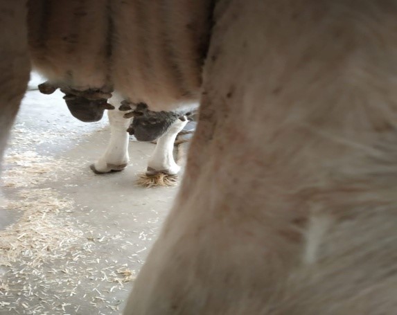
Bovine papillomatosis is a viral disease of cattle caused by bovine papilloma viruses (BPV), belonging to genus Papilloma virus, family Papillomavaviridae and manifested as benign tumors or warts on skin surface. Usually it appears as multiple, sessile or pedunculated, circumscribed grey white to dark brownish black outgrowth on skin over different body parts and may be smooth surfaced, spherical or horny (Singh et al., 2009). The lesions are commonly seen on teats and udder and can also appear on other parts like head, neck, around eyes, shoulder region and ventral abdomen (Sharma et al., 2004). In cattle, thirteen types of BPV have been identified (De Villiers et al., 2004), among which BPV type 1-11 can cause teat warts in dairy cattle (Dagalp et al., 2017). The presence of warts on teats of lactating cows can create severe milking problems and make them susceptible to secondary bacterial invasion resulting in mastitis and lowered milk production. The presence of warts on teats and udder also reduces animal cost.
Etiology
In cattle, bovine papilloma virus (BPV) is the main etiological agent of cutaneous and teat papillomatosis (Campo, 2002) and produces characteristic gross lesions that are either proliferating outwards or inverted and are composed of hyperplastic epithelium supported by discrete dermal tissue containing dilated capillaries (Hargis et al., 2012). Papillomaviruses (PVs) are members of the Papillomaviridae family, which have a characteristic circular double-stranded DNA genome of around eight kilobase pairs (kbp) that usually contains at least six relatively conserved open reading frames (ORFs) in an early (E1, E2, E6, E7) and a late (L1, L2) region.4 Papillomaviruses are characterized genetically by the L1 ORF.4 To date, at least 112 nonhuman papillomaviruses have been identified, and there is the anticipation that more will be detected.1 It appears that each species carries a suite of papillomaviruses- for example, 13 bovine papillomavirus (BPV) types have been identified in cattle (BPV-1 through BVP-13), 15 canine papillomavirus (CPV) types in dogs (CPV-1 through CPV- 15), and 7 equine caballus papillomavirus (EcPV) types in horses (EcPV-1 through EcPV-7). The feline sarcoid-associated virus has been proposed as BPV-14; it can infect cattle but has not been demonstrated in horses.
Cattle types show some site predilection or site specificity, as exemplified by the following partial listing:
Epidemiology
Papillomatosis has an international occurrence in all animal species. BPV-2 infection is common in horses and cattle. The method of spread is by direct contact with infected animals, with infection gaining entry through cutaneous abrasions. Viruses can also persist on inanimate objects in livestock buildings and infect animals rubbing against them. All species may be affected by papillomas or fibropapilloma, but it is most commonly reported in cattle and horses. With cattle, usually several animals in an age group are affected. Alimentary papillomas occur in up to 20% of cattle ingesting bracken fern. Papillomavirus infection is widespread in nondomestic species, including birds and reptiles, and is associated with disease in many of these species. Cutaneous papillomas of the head and neck occur predominantly in young animals. The occurrence of cutaneous warts and their severity can be influenced by factors that induce immunosuppression, and latent infection has converted to clinical disease with the administration of immunosuppressive agents.
Clinical signs
The most common papillomas occur in the skin of cattle under 2 years of age, most commonly on the head (Fig. 16-2), especially around the eyes, and on the neck and shoulders (Fig. 16-3), but they may spread to other parts of the body. They vary in size from 1 cm upward, and a dry, horny, cauliflower- like appearance is characteristic. In most animals they regress spontaneously, but the warts may persist for 5 to 6 months, and in some cases for as long as 36 months, with serious loss of body condition. Warts on the teats manifest with different forms depending on the papillomavirus type and may show an increasing frequency with age. The frond forms have filiform projections on them and appear to have been drawn out into an elongated shape of about 1 cm in length by milking machine action. If sharp traction is used, they can often be pulled out by the roots. The second form is the flat, round type, which is usually multiple, always sessile, and up to 2 cm in diameter. The third form has an elongated structure appearing like a rice grain. Teat warts may regress during the dry period and recur with the next lactation. Perianal warts are esthetically unattractive, but they do not appear to reduce activity or productivity. Genital warts on the vulva and penis make mating impracticable because the lesions are of large size, are friable, and bleed easily. They commonly become infected and flyblown. They occur on the shaft or on the glans of the penis in young bulls, may be single or multiple, are pedunculated, and frequently regress spontaneously.
Diagnosis
Cases of papilloma can be done on the basis of visible lesion present on skin of different part of the body.
Differential diagnosis
Clinically, there is little difficulty in making a diagnosis of dermal papillomatosis, with the possible exception of atypical papillomas of cattle, probably associated with an unidentified type of the papillomavirus. These lesions are characterized by an absence of dermal fibroplasia and are true papillomas rather than fibropapillomas. All ages of animals can be affected, and the lesions persist for long periods. They are characteristically discrete, low, flat, and circular, and they often coalesce to form large masses. They do not protrude like regular warts, and the external fronds are much finer and more delicate.
Treatment
Warts can be removed by surgery or cryosurgery. Crushing of a proportion of small warts, or the surgical removal of a few warts, has been advocated as a method of hastening regression, but the tendency for spontaneous these treatments very difficult. Surgical removal can be followed by vaccination with an autogenous vaccine, although the efficacy of this approach is unclear. There is anecdotal concern that surgical intervention, and even vaccination, in the early stages of wart development may increase the size of residual warts and prolong the course of the disease. Aural plaques in horses can be treated by application of imiquimod, an immunomodulator and antiviral agents, as 5% cream applied three times a week, every other week, for 6 weeks to 8 months. Crusts were removed before each application of the cream and required sedation in most horses.
Prevention and control
Specific control procedures are usually not instituted or warranted because of the unpredictable nature of the disease and its minor economic importance. Vaccination has been shown experimentally to be an effective prevention method and gives complete protection in cattle against stiff experimental challenge. The vaccine must contain all serotypes of the papillomavirus because they are very type-specific. Avoidance of close contact between infected and uninfected animals should be encouraged, and the use of communal equipment between affected and unaffected animals should be avoided.
Vaccination
For cattle, autogenous vaccines prepared from wart tissues of the affected animal are effective in many cases. Commercially available vaccines are available for cattle but may be less efficacious; an autogenous vaccine prepared for a specific problem has the advantage of including the local virus types. The vaccine is prepared from homogenized wart tissue that is filtered and inactivated with formalin. Because of the different BPV types, care is required in the selection of the tissues. In general terms they can be selected based on tumor type, location, and histologic composition. The alternative is to use many types of tissue in the vaccine. Animal to animal variation in regression following vaccination of a group of calves with a vaccine prepared from a single calf in the group has been attributed to more than one BPV type producing disease in the group. The stage of development is also important, and the virus is present in much greater concentration in the epithelial tissue of older warts than young ones. The vaccine can be administered subcutaneously, but better results are claimed for ID injection. Dosing regimens vary, but 2 to 4 injections 1 to 2 weeks apart are commonly recommended. Recovery in 3 to 6 weeks is recorded in 80% to 85% of cases where the warts are on the body surface or penis of cattle, but in only 33% when the warts are on the teats. The response of low, flat, sessile warts to vaccination is poor. Development of DNA vaccines for prophylaxis or therapy (which will likely use different genes) is active but experimental at this time.
References
- Munday JS. Bovine and human papillomaviruses: a comparative review. Vet Pathol. 2014;51:1063-1075.
- Rector A, van Ranst M. Animal papillomaviruses. Virology. 2013;445:213-223.
- Dairyman, H. (1993) Herd Health, W.D. Hoard & Sons Company, USA.
- Hagan WA, Bruner DW, Timoney JF (1988) Hagan and Bruner’s Microbiology and Infectious Diseases of Domestic Animals (8th Edn), Cornell University Press,
- Divers TJ, Peek SF (2008) Rebhun’s Diseases of Dairy Cattle. Saunders Elsevier, USA.
- McKenna J (1998) Natural Alternatives to Antibiotics, Avery Publishing group, USA.
- Grey, Hampel (1847) The Homeopathic Examiner vol II, Leavitt, Tsow & Co., Printers.
- Macleod G (2012) The Treatment of Cattle by Homoeopathy, The C.W. Daniel publisher, England.


