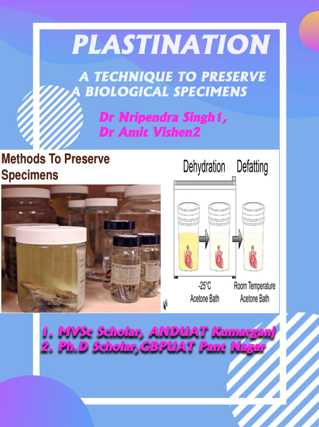
PLASTINATION- A TECHNIQUE TO PRESERVE A BIOLOGICAL SPECIMENS
Plastination is the process of tissue preservation by embedding tissue with synthetic polymer like Silicon,Polyester ,Epoxy, Resin etc. to Produce a Dry, Durable, Handy and Natural looking Specimen useful as a unique tool for long term Educational Purpose.This method was invented in 1978 at the university of Heidelberg by Dr. Gunther von Hagens .
THE PRINCIPLE BEHIND THE TECHNIQUE
This technique can be used to preserve biological specimens that involve replacing water and fat in tissue with a polymer. This method produces “ Plastic “ Bodies or organs which are :- Non Toxic, Odourless , Dry and Durable, Can be handled very easily for Examination & Long lasting. Among the Polymers , the commonly used are Epoxy, Silicone and Polyester
For obtaining the Best Plastinated Specimens , The Polymer used must have the following Desirable Properties:-
- 1. Lower possible viscosity in uncured state .
- 2. Its refractive index of the polymer should be different from that of tissue or else a transparent specimen would be obtained.
- 3. Curing should not be inhibited by the tissue
- 4. Mechanical properties of the polymer should be appropriate when cured i.e, it should be rubber like to stimulate a natural state or firm
- 5. It should be Affordable.
STEPS IN PROCESSING THE SPECIMEN FOR PLASTINATION
STEP 1 – FIXATION
- The specimen should be fixed in 10% formalin. This stabilises the tissues and prevent autolysis.
- To enhance colour presrvation cold kaiserling solution containing 5% formalin should be used.
- Specimen is generally fixed by injection through blood vessels ( If not previously filled with colouring material)
- By infiltration i.e., injection of the solution in the muscles with the help of a syringe and needle .
- By immersion in the fixative solution.
- In case of specimen stored in solution containing glycerol,these specimen have to be rinsed off thorougly to remove all the glycerol before being Plastinated.
STEP 2- DEHYDRATION
- Different types of polymer used for Plastination are not miscible with water hence the specimens must be absolutely dehydrated.
- Their water must be replaced by an intermediary solvent to permit penetration of the polymer with the specimens
- Dehydration can be achieved in stepwise ethanol baths , but the standard procedure is freeze substitution with acetone at -25°c.
- When immersed into cold acetone the specimens freeze immediately and shrinkage is considerably reduced .
- This procedure takes 4 to 5 weeks , with 3 changes of acetone
- The lipid rich specimen now have to be transferred in acetone at room temperature for one week to achieve Defattening and then they can be impregnated.
- Specimen prepared for Epoxy Plastination needs extra Defattening Bath in Methylene chloride to improve their transparency
STEP 3- IMPREGNATION
- This is performed in a vacuum chamber where the acetone saturated specimens are submerged into a bath of liquid polymer.
- With a vacuum pump the pressure is slowly decreased in the chamber . The acetone is the changed from its liquid phase to vapour phase and aspirated by the vacuum pump. The extraction of the acetone creates a vacuum inside the specimen that forces the penetration of polymer into them, down to their microscopic level
- Because of the great difference between the high vapour pressure of the acetone and low vapour pressure of the polymer, only the Acetone is extracted when the vacuum is applied
STEP 4- HARDENING & CURING
- Finally the polymer inside the specimen has to be Cured/Hardened
- This is achived by exposing the impregnated specimen to a hardener which can be liquid or gases in nature
- The impregnated specimen and a bowl filled with curing agent is placed in a tightly closed chamber for several week
- To enhance the curing procedure air may be bubbled through the fluid
- For Complete curing the specimen should be kept in a plastic bag for several week
TYPES OF PLASTINATION
On the basis of size, shape and nature of tissue, there are three types of plastination viz. Whole body/organ plastnation, Luminal cast plastination and Sheet plastination
- Whole organ or a body Plastination- in this method, Silicon (S10) and polypropylene resins are used. Using this technique, whole of the structure or organ, and its relationships can be preserved
- Luminal cast plastination- is done for hollow organs like lungs, stomach, intestine, ventricles of brain, vascular pattern of heart and kidneys. Specimens are dilated/inflated during fixation, dehydration and curing. Beautiful and precise bronchial pattern can be seen by this technique.
- Sheet plastination – In this method, thin transparent or thick opaque sections of body or an organ are preserved. These sheets are portable and shows cross sectional anatomy of organs equivalent to CT or MRI scan sections. Sheets can be taken in various planes. Thin sections (1-2mm) of organs are similar to routine histology slides. Polymers such as epoxy (E12), polyester (P35) or polypropylene (araldite) resins are used for making sheet plastinates.
ADVANTAGE OF PLASTINATION
- The Specimen are Dry, Easy to handle, Store, Transport and long lasting.
- Non Hazardous, Non Infectious, doesn’t radiate fumes or fluids
- Can be used for imperative preparation of specimens for Museum Display.
- Can be used for Preparation of samples for Evidence
- Storage and Maintainence is easy. They can even be stored in Plastic Bags with Essential Credentials.
DISADVANTAGE OF PLASTINATION
- Costly Procedure
- Time consuming
- Required skill technical support to carry out the procedure and in handling the equipmemts.
- Prepared specimen requires handling with care.
- Chemical used such as Acetone are highly inflammable and should be used in place equipped with fire extinguishing measure.
- https://www.pashudhanpraharee.com/plastination-a-favourable-approach-to-preserve-biological-specimens/
- Dr Nripendra Singh1, Dr Amit Vishen2
- MVSc Scholar, Department of Veterinary Anatomy & Histology College of Veterinary Science & Animal Husbandry ANDUAT Kumarganj Ayodhya 224229
- D Scholar, Department of Veterinary Anatomy & Histology College of Veterinary Science & Animal Science GBPUAT Pant Nagar Uttrakhand 263145
- Reference-http://www.vhlab.umn.edu/atlas/methodologies/preservation/plastination.shtml#:~:text=Plastination%20is%20a%20preservation%20method,this%20method%20in%20the%201970s1


