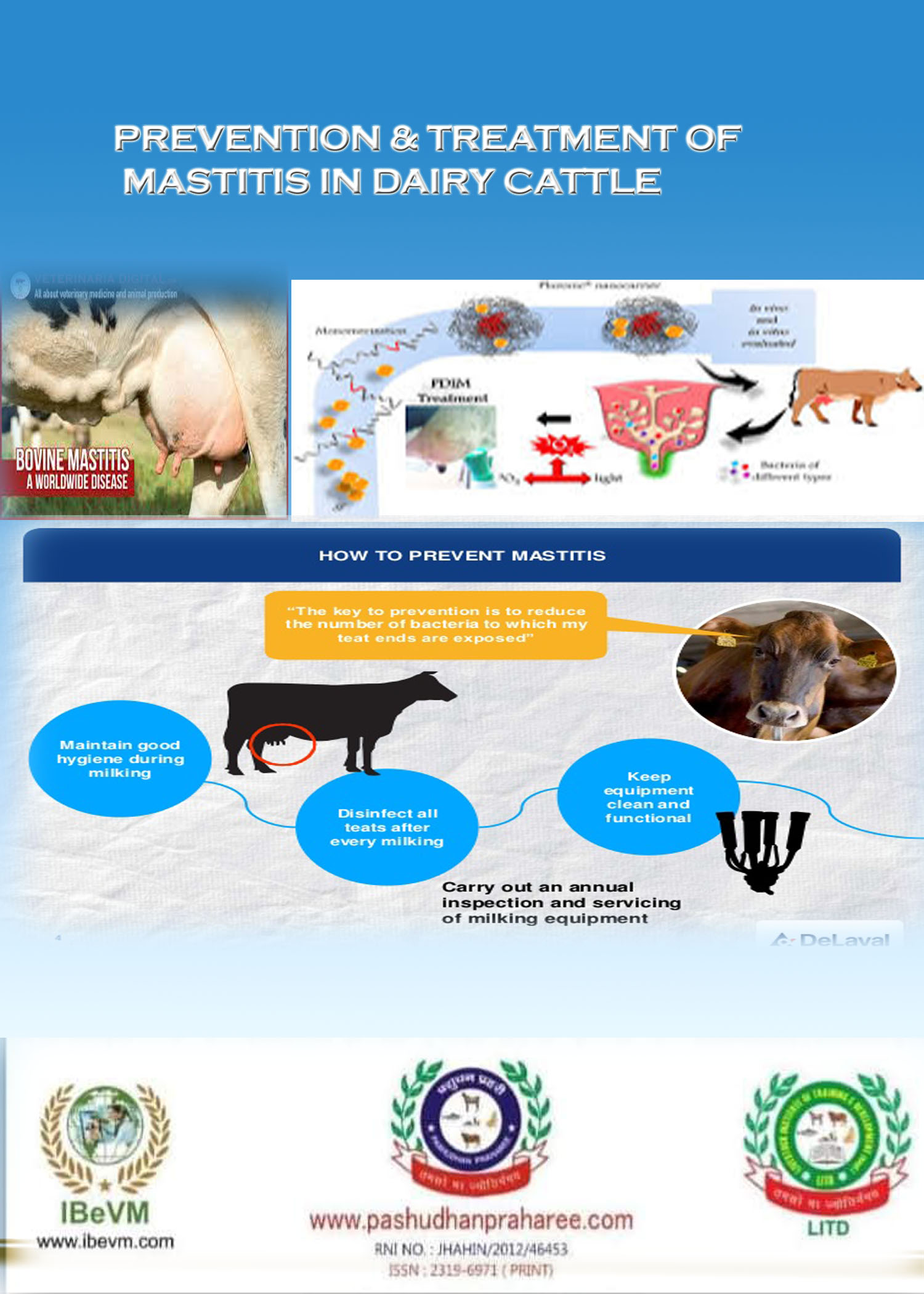
PREVENTION & TREATMENT OF MASTITIS IN DAIRY CATTLE
Post no-589 Dt-04/03/2018

Compiled & edited by-DR RAJESH KUMAR SINGH, JAMSHEDPUR, 9431309542,rajeshsinghvet@gmail.com
Mastitis is inflammation of the mammary gland usually caused by pathogens, mainly
bacteria which have entered the teat canal. Intramammary infection usually
occurs as an immune response of animal to bacterial invasion to eliminate invading
pathogen. Bacterial presence within the udder results in the movement of white blood cells
into the gland to help ght the disease. An uninfected mammary gland will maintain a low
somatic cell count (<200,000 cells/ml of milk). Once gland tissue becomes infected,
numerous neutrophils will be drawn to the mammary gland, resulting in increased somatic
cell counts.
Types of mastitis
A- Contagious Mastitis: Caused by bacteria live on the skin of the teat and inside the udder. Contagious mastitis can be transmitted from one cow to another during milking.
B- Environmental mastitis: Describes mastitis caused by organisms such as Escherichia coli which do not normally live on the skin or in the udder but which enter the teat canal when the cow comes in contact with a contaminated environment. The pathogens normally found in feces bedding materials, and feed. Cases of environmental mastitis rarely exceed 10% of the total mastitis cases in the herd.
Contagious mastitis can be divided into three groups:
1- Clinical mastitis
2- Sub-clinical mastitis
3- Chronic mastitis
Clinical mastitis
Characterized by the presence of gross inflammation signs (swelling, heat, redness,pains). Three types of clinical mastitis exist.
1.1- Peracute mastitis
Characterized by gross inflammation, disrupted functions (reduction in milk yield, changes in milk composition) and systemic signs (fever, depression, shivering, loss of appetite and loss of weight).
1.2- Acute mastitis
Similar to percute mastitis, but with lesser systemic signs (fever and mild depression).
1.3- Sub-acute mastitis
In this type of mastitis, the mammary gland inflammation signs are minimal and no visible systemic signs.
2- Sub-clinical mastitis
This form of mastitis is characterized by change in milk composition with no signs of gross inflammation or milk abnormalities. Changes in milk composition can be detected by california mastitis test
3-Chronic mastitis
An inflammatory process that exists for months, and may continue from one lactation to another. Chronic mastitis for the most part exist as sub-clinical but may exhibi periodical flare-ups sub-acute or acute form, which last for a short period of time.
Type Contagious mastitis Environmental Mastitis
Source of Infection Teat and udder Contaminated environment
Transfer of infection
in to udder During Milking Between milking and during
dry period
Clinical mastitis Most cases are subclinical Higher proportion are clinical
Control by Post milking teat dipping, dry
cow therapy, milking hygiene
and culling of chronic cases Environmental hygiene,
predipping, dry period
teat sealant
Factors influencing susceptibility to mastitis—-
1. Type of bacteria: Some bacteria are more virulent than others in causing mastitis.
2.Physiological status of cow: Although infection can occurs at any time, most of the new infections take place during the first three weeks of the dry period and during the first month after parturition, suggesting that level of milk production is not directly related tomastitis. It is likely that intramammary pressure is a predisposing factor for mastitis during these periods.
3. Age of the cow: The incidence of mastitis increases with age. Nevertheless, it is possible
for the udder of the first-calf heifer to be infected at parturition.
4. Level of milk production: Not directly related to incidence of mastitis. However, other
factors, which affect milk, yield such as milking rates, pendulous udders may be related
to mastitis incidence.
5. Inherited features of the cow: Length of the leg in proportion to the udder size and relative strength of the udder attachment are examples. Large, pendulous udders tend to exceed the capacity of the supporting ligaments, with a consequent of breakdown of the udder. This will subject the udder to more physical injuries and thus increases the incidence of mastitis.
6. Milking machine: Improper use of milking machine (irregular fluctuation of vacuum level, over-milking, incomplete milking) is related to tissue irritations and incidence of mastitis.
7. Environment: Mastitis often increases when cows are turned onto pastures. Chilling of the udder in cold ground in the spring or fall. Housing as it relates to the degree of udder and teat injury.
Principles of controlling mastitis: —-
1. Elimination of existing infections in udder: Antimicrobial therapy during dry period
is a one of the method of choice. Dry period antibiotic treatment is a formulation of
antibiotics prepared for administration into the udder immediately after the last milking of
lactation to reduce new infections and elimination of existing infections. In India, ceftiofur
hydrochloride 500mg/10ml preparation is available for the treatment of sublinical mastitis at
the time of dry off mainly associated with Staphylococcus aureus, Streptococcus dysgalactiae
and Streptococcus uberis. There are three phases of dry period namely early, mid and late dry
period. In the first two weeks (early dry period); keratin plug forms a teat seal and active
involution of udder tissue occurs. In the mid dry period, immunoglobulins and natural
inhibitory substances like neutrophils and lactoferrins are formed. In the last two weeks
before calving (late dry period), keratin plug slowly dissolves and colostrogenesis occurs.
Animals are most succeptible to infection during first and last two weeks of dry period
because teat plug is forming and dissolving respectively. Further depressed immune system
of dam with approaching parturition contributes to new infections. The persisted subclinical
infections during dry period may are into clinical cases after calving. Most new cases of
mastitis occur first four weeks of lactation and 60% of clinical cases by environmental
pathogens originate from the infections established during early and late dry period .
2. Prevention of new infection: Premilking and postmilking teat disinfection are the
most effective mastitis control practice in lactating animals. Premilking teat disinfection
with chlorhexidine in association with post milking teat disinfection reduces new
intramammary infection. Post milking teat disinfection is regarded as the single most
effective control practice in lactating animals. Iodophores teat dips with 0.1 to 1% available
iodine can be used as post dip. Post milking teat disinfection removes bacteria deposited
during the milking process and therefore it is an extremely important control measure against
contagious mastitis. Postdip should be applied as soon as milking is over. Teat must not be
wiped dry after post dip. Premilking teat disinfection is aimed at reducing the incidence of
environmental mastitis.
3. Monitoring udder health status: Implementing and effective system of monitoring udder health involves monitoring at herd and individual level. Use of animal side diagnostic test like California mastitis test – somatic cell count and milk bacteriological culturing are important for udder health and milk quality.
Five points mastitis control programme promoted by The National Mastitis Council (NMC) is as follows:
1. Udder hygiene and milking management
2. Milking equipments maintenance
3. Dry animal therapy
4. Appropriate therapy of mastitis during lactation
5. Culling chronically infected animals
Udder hygiene includes premilking udder preparation by washing teat in water and drying of teat with paper towel followed by use of 0.25% iodine premilking teat disinfectant (Dipping is better than spraying).
The foremilk stripping is checked for clinical mastitis (clot, watery or stringy milk) using a strip cup test. The early recognition and treatment of clinical cases is important part of mastitis control programme (Radostits, 2000).
Following package of practices is very useful for the control of mastitis in dairy herds.—-
Adoption of proper hygienic measures:
Maintenance of proper hygiene is perhaps the most important management practice in mastitis control as it affects the degree of exposure and population of microbes in the environment surrounding the cow.
The sanitary measures can be summarised as follows:
1. Teat washing with disinfectant solution and wiping with individual clean towels, prior to each milking.
2. Disinfecting of hands of milkers, milking machine clusters before milking and
3. Teat dis-infection after each milking by dipping or spraying all teats in disinfectant solution.
Suitable udder disinfectants for this purpose are as follows:
• Iodophor solution containing 0.1 to 1.0% available iodine.
• Chlorhexidine 0.5 or 1% in polyvinyl pyrrolidone solution or as 0.3% aqueous solution
• Sodium hypochlorite (4% solution).
To ensure effectiveness of these disinfectants the udder must be washed clearly to remove all the organic matter before applying disinfectant solution on them.
Proper milking procedure:
Proper milking of dairy animals is important regardless of whether hand or machine milking is being followed. Rapid and full hand milking is desirable as this ensures harvesting of more milk and simultaneously prevents teat injury which might result as a consequence of improper milking method (Fisting etc). Milking management becomes more important when machine milking is followed. In addition to proper disinfecting of milking machine, the following factors must be considered.
• Check fore milk and udder for mastitis using strip cup or California mastitis test (CMT).
• Attach the milking unit properly.
• Optimum vacuum must be ensured.
• Pulsation rate should be maintained within permissible limits.
• Teat liner must be checked for rupture etc. and must be replaced at least once a month.
• Milk the infected animal in the end when all the fresh animals have been milked.
• Inspect milking equipment routinely.
Dry cow therapy:
The dry period offers a valuable opportunity to improve udder health while cows are not lactating. On the other hand, the beginning (initial 2 3 weeks) and the final 2 3 weeks of gestation period is very vulnerable to new infections. The procedure of dry cow therapy may be carried out as follows:
• Dip all the teats in an effective teat dip after complete milking and dry completely.
• Disinfect each teat end with alcohol soaked cotton swab and infuse a single doses x syringe of a recommended antibiotic. Long acting antibiotic preparations like benzathine Cephapirin, benzathine cloxacillin, benzathine penicillin, erythomycin, novobiocin etc, can be used successfully. A partial insertion method of administration is better than complete insertion.
• Immediately after treatment dip all the teats in an effective teat dip again.
Quick diagnosis and appropriate therapy of affected animals during lactation.
Early detection of mastitis, preferably in sub-clinical form itself, is the key to the successful treatment of the disease. This can be better done by screening all quarter samples using California Mastitis Test (CMT) and monitoring Somatic Cell Count (SCC) at least once a month regularly. Use of strip cup is another easy test for detecting clinical mastitis. The antibiotic therapy should be done after conducting the sensitivity test and use single dose tubes and not the multiple dose bottles which can become contaminated. It is better to consult a qualified veterinarian.
Segregation and culling of chronic infected animals:
As soon as the mastitis is confirmed, the cow must be segregated from rest of the herd and milked and treated separately besides adopting proper hygienic measures. Selective culling of the cows with chronic mastitis (three or more episodes in lactation) should be practised.
Monitoring udder health status:
• Monitoring udder health of individual cows individual cow SCC can be used along with CMT. For proper monitoring good record keeping is essential. Based on the current udder health status the appropriate control measures should be undertaken. Besides, periodic review of the udder health management programme is also important to take corrective measures wherever required.
Setting goals for udder health status:
Establishment of realistic periodic targets for various udder health parameters is the final step of a complete udder health management program. The goals should be realistic. To sum up, the production of clean milk requires looking at and evaluating nearly every aspect of the milk production system. To consistently produce high quality milk with low bacteria counts requires continual attention to numerous details. You should not be satisfied with any other product or equipment on your farm that just barely met minimum performance standards.
HOMEOPATHY MEDICINE OF MASTITIS—
(BY DR. Sawapan Kumar Dey sir)–
. Mastitis & Fibrosis (cattle).
For Mastitis Phytolacca 200 + Hepar sulph 200 + Belladona 200 mixing in globules 40 Dose 5 globules twice daily until recovery.
For Fribrosis Conium 200 + Calcaria flour 200 + Graphitis 200 mixing in globules 40 Dose 5 globules twice daily until recovery
No milk after delivery (cattle)
1) Phytolacca 3x. Dose 10 drops twice daily.
2) Urtica urens 30 mixing in globules 40 Dose 5 globules thrice daily.
** Continue until satisfactory result.
Increase or stop milk production(cattle)
To increase production: phytolacca 3x Dose 10 drops twice daily for 10 days.
To stop production during last stage of pregnancy: Lac caninum 1m mixing in globules 40 Dose 5 globules once daily for 7 days.
Blood in milk (cattle) :
Ipecac 200 + Bufo 200 (combined) mixed with globules size 40. Five globules twice daily
Therapeutic management: —
Mastitis is the most frequent cause of antibacterial use on dairy farms and contributes to a
substantial portion of total drug and veterinary costs incurred by the dairy industry . Knowledge on the microbiological prole of mastitis by clinical and laboratory
diagnosis (isolation, typication and antibiogram) is one of the basic pillars for the rational
use of antimicrobial agents (ATM).The aim in selecting the best antimicrobial treatment
regimen for mastitis is administering the drug at a dose and site that will allow accumulation
in the mammary gland to maintain an effective drug concentration. There are three
pharmacological compartments. The most common target compartment milk and epithelial
lining of duct and alveoli of mammary gland. The S. agalactiae, S. dysgalactia, Coagulase
negative staphylococci pathogen tend to locate in duct areas of the udder where antibiotics
are effective S. agalactiae are very sensitive to penicillin, so treatment has a relatively high
cure rate by administering intramammary administration . Second
compartment is deep tissue of the mammary gland. S. aureus can penetrate into udder tissue
and form micro abscesses that are protected from antimicrobials by scar tissue. They are
difcult to cure, especially during lactation, so prevention is essential. Coliform mastitis
involve animal itself, third compartment. Bacteraemia can occur and respond to systemic
therapy. Mild cases of clinical coliforms mastitis generally are self-limiting and the animals
own defense mechanisms can successfully clear the infection from the udder, and though
antibiotics are not required at all; serious cases supports the use of systemic administration of
antibiotics. Pseudomonas infection is impossible to treat so animal must be culled. In
mastitis caused by penicillin-susceptible S. aureus strains, best results were achieved using a
combination of systemic and intramammary treatment with penicillin G. In infections of the
milk compartment such as streptococcal mastitis, there is probably no advantage of systemic
administration indeed the concentration of penicillin G in milk remains 100-1000 fold lower
than when given intramammarily.
Treatment
Antibiotic treatment: Typically when clinical mastitis is detected, the cow is milked out and then given an intramammary infusion of antibiotic, ie. infused directly into the infected gland.
Intramammary infusion technique
Apply antibiotics directly into the udder. The intra mammary infusion composed of antibiotics can be infused through teat canal,The intra mammary infusion are available in the market made by different companies with different brand names, generally consist of combinations of antibiotics like Streptomycin, Neomycin, Erythromycin, Polymixin etc
How to apply antibiotic directly into the teat:
Step 1: Milk the udder until it is empty
Step 2: Clean the end of the teat
Step 3: Put the tip of the tube into the teat and squeeze the antibiotic up into the udder
Step 4: Massage the teat and the udder
If the disease is severe, also give antibiotics by injection!
The following drugs can be used
Antibiotic composed of ceftriazone plus sulbactum or amoxicillin plus sulbactum or ceftizoxime along with anti inflammatory drug for 3-5 days under the supervision of vet.
Reasons for treatment failure : —-
Reasons for treatment failure include lack of contact between bacteria and antibiotics due to scar tissue formation, protection within leukocytes, poor drug diffusion, and inactivation by milk and tissue proteins; microbial resistance to antibiotics; improper treatment procedures like stopping the therapy too soon.
reference-on request


