

(1)")
Surgico-therapeutic management of ruminal impaction in Patera Buck: A case report
Jagmohan Rajpoot1, A.S. Parihar2, Megha Gaur3*
1M.V.Sc. Scholar, 2Assistant Professor, 3M.V.Sc. Scholar
*Corresponding author: Dr. Megha Gaur, Mob. No. 8770278093, E-mail ID- vetmeghagaur1996@gmail.com, Postal address- H. No. 181, ward- 13, Kamad road, Indergarh, Dist.-Datia (M.P.) – 475675
Abstract
A 1.6 years old Patera breed buck was presented to Veterinary Clincal Complex, College of Veterinary Science and Animal Husbandry, Mhow with the history of absence of rumination since 6 days and recurrent bloat. Abdominal palpation revealed ruminal impaction for which exploratory lapro-rumenotomy was performed under standing condition by paravertebral regional anesthesia and local infiltration with lignocaine hydrochloride 2% solution over the site of incision. Approx. 8 kg of ruminal content was removed. The suturing was done in regular manner (rumen, peritonium, fascia and skin). Fluid was administered during surgery and after surgery. The buck started rumination after 5 days of surgery and recovered without complications.
Key words
Regional, rumenotomy, paravertebral and buck
Introduction
Local, regional, and spinal anesthesias are safe, effective, often more desirable procedures for ruminants than general anesthesia. However, rumenotomies are a common practice for the removal of foreign bodies in goats (Hayder et al., 2006), and this technique is also used for zootechnical or research purposes (Martinez et al., 2019). Although most of the diseases affecting the gastroenteric tract in ruminants are managed medically, some require surgical treatment (Radostits et al., 2007). In this case, successful recovery of rumination in buck was noted after the surgery.
Case history and observation
A 1.6 years old buck, weighing 80 kg was presented to Veterinary Clincal Complex, College of Veterinary Science and Animal Husbandry, Mhow with the history of absence of rumination and recurrent bloat since 6 days as it was kept in room with warm environment. The buck was anorectic and urine was dark yellow in colour. Temperature and respiratory rate were normal. Abdominal palpation of buck revealed ruminal impaction for which exploratory lapro-rumenotomy was performed.
Treatment and discussion
The buck was prepared for surgery in routine manner. The surgery was performed in standing position by regional anesthesia (paravertebral) and local infiltration around the incision site of the operation with 2% lignocaine hydrochloride. Intravenous fluid was administered prior to surgery and during surgery (RL, and NS). Antibiotic Ceftriaxone tazobactam 1gm and analgesic Meloxicam @.5mg per kg bt wt were administered intravenously prior to surgery. A vertical incision was given aspetically just behind the last rib, and about three centimeters from the transverse lumbar process (Dharmaceelan et al.,2017), just above the dorsal sac of the rumen (Lozier and Niehaus, 2016). The incision was extended over the skin, fascia, peritonium and rumen. As the rumen got exposed, the rumen wall was fixed with skin in order to prevent the content (ruminal) into the peritonium. The ruminal content was removed out about 8kg paste like. After removal of content flushing was done by metronidazole and normal saline about 300ml and 2 litre respectively. Suturing of rumen wall was done by simple continuous manner with catgut no. 1. Peritoneum was sutured in regular manner with absorbale suture vicryl no 1. Further skin suturing was done by simple cross mattress pattern with nylon no. 1. Oint. betadine was applied over the suture line and dressing was done. Post operatively buck was maintained on intravenous fluid 4 litre RL and 300ml metronidazole was given slow intravenous for 2 days and antibiotic and analgesic were continued for 3 days. Along with that Inj. Pheniramine maleate @ .3mg per kg bt wt and Inj. Trivibet 2 ml were also given intramuscularly. Daily dressing was done with oint. betadine, oint. himax and topicure spary in order to avoid postoperative complications and promote healing. The skin got healed 5 days postoperatively and sutures were removed after 12 days of surgery. The buck got relief after 3 days of rumenotomy untill it keeps on fluid therapy and on 4th day it starts rumination. The buck started rumination after 4 days and regain its appetite after 2 weeks of surgery.
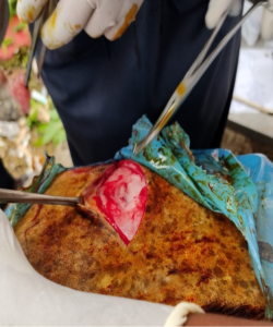

Fig 1. Vertical incision just behind last rib Fig. 2 Exteriorozation of dorsal sac of rumen

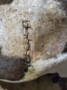
Fig. 3 Fixation of ruminal wall to skin with stay suture Fig. 4 Skin sutures postoperatively

Fig.5 Healed skin after 12 days of surgery
References
Adamu. SSS. Zira. GII. Egwu. GOO. Dilli. HKK. (1993). A simplified polythene drape technique for reducing post-rumenotomy complications in goats. Small Ruminant Research. 9(4):389–394.
Baillie. S. Anzuino. K. (2006). Hairballs as a cause of anorexia in angora goats. Goat Veterinary Society Journal. 22(January 2006):53–55.
Das. J. Behera. SS. (2011). Acute bloat in a goat and its surgical management by rumenotomy. Intas Polivet. 12(II):322–324.
Dharmaceelan. S. Kumaresan. A. Kanjana. D. (2017). Surgical management of Ruminal Impaction in a goat. Intas Polivet. 18:329–330.
Edmondson. MA. (2016). Local, regional, and spinal anesthesia in ruminants. Veterinary Clinics of North America: Food Animal Practice. 32(3):535–552.