Treatment of Guttural Pouch Tympany in Horses
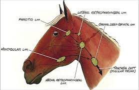
The guttural pouches are only found in horses and their cousins, such as zebras and donkeys. The guttural pouches are rather splendid outpouchings of the eustachian tubes, the connection between the middle ear and the back of the oral cavity responsible for equilibrating pressures between the ear and our outside surroundings.
The gutteral pouches are framed by the base of the skull at the top, the pharynx and esophagus at the bottom, and the salivary glands and mandible on the sides. There is a slit-like opening from each gutteral pouch into the pharynx that is hard to see, and even harder to open for examination or drainage purposes.
Both guttural pouches are divided into two separate compartments by the stylohyoid bone. The one closer to the body’s midline, called the medial compartment, contains many different nerves and arteries, including the internal and external carotid arteries, five cranial nerves, a few lymph nodes and some delicate bones and joints. This makes diagnosis and treatment of gutteral pouch problems difficult.
The guttural pouches are lined with the same respiratory epithelium, or tissue, that lines the upper airway. This means that the guttural pouch can be affected by viruses and infections that affect other portions of the upper airway.
In the horse, the guttural pouch can be seen only by endoscopic examination. Two flaps, one on each side, are visible just before the voicebox (larynx). These flaps ordinarily do not allow any air into the guttural pouches.
Guttural pouch tympany may be present in one or both pouches. Clinical signs of GPT may be seen as early as hours after the foal is born. Research show that GPT effect more female foals more than males. Arabian and Hanoverian breeds are more predisposed to GPT. Guttural pouch tympany can also be found in other breeds such as the American Saddle Horse, Quarter Horse, Appaloosa and the English Thoroughbred.
Guttural pouches are located on both sides of the foal’s head near the upper mandibles; they are mucosal sacs of the Eustachian tubes (auditory tubes). Guttural pouch tympany is a condition where air flows through the ear canal and then gets trapped inside the guttural pouch. As more air gets trapped in the guttural pouches, the pouches enlarge, which then cause the pharynx and larynx to be compressed.
Symptoms of Guttural Pouch Tympany in Horses
Symptoms may include one or more of the following:
- Swelling in the cheeks
- Swallowing problems
- Labored breathing
- Distended nostrils
- Milk discharged through the nostrils
- Difficulty nursing
- Fever
- Cough
- Edema
- Respiratory infections
- Pneumonia
Causes of Guttural Pouch Tympany in Horses
Guttural pouch tympany is a congenital disease in horses, which means the foal is born with GPT. The exact cause of guttural pouch tympany is unknown. Horses diagnosed with guttural pouch tympany should not be allowed to breed because they would be passing on the genetic condition to the offspring.
Diagnosis of Guttural Pouch Tympany in Horses
Diagnostic tests included in the evaluation of your foal may include:
- Complete blood count – Checks the count of platelets, red and white blood cells; this helps determine if there is a bacterial infection
- Fecal exam – Can help diagnose parasites and if there is any blood in the feces
- Urinalysis – Checks for kidney function, crystals, blood or bacteria in the urine
A veterinarian will be able to diagnose the guttural pouch tympany by a physical examination of your horse. Diagnostic testing may be recommended to confirm the diagnosis and to make sure there are no secondary conditions. The veterinarian will recommend x-rays of the foal’s head. If the veterinary doctor heard fluid in the foal’s lungs, chest x-rays may also be suggested. The veterinarian may also choose to perform an endoscopic examination. The endoscope is inserted through the nostril and moves along the respiratory tract. The procedure allows the veterinarian to have a visual of the guttural pouch. The foal will need to be sedated for this procedure.
Treatment of Guttural Pouch Tympany in Horses
Once guttural pouch tympany is diagnosed, the veterinarian may insert a temporary catheter into the pouch to release the trapped air. This will give the foal some relief from the pressure in his airway. He will also be able to nurse normally. If a bacterial infection was found, the veterinarian will prescribe antibiotics. He may also recommend a non-steroidal anti-inflammatory, analgesic drug such as flunixin.
The permanent solution for guttural pouch tympany is surgery to correct the defect. The equine veterinarian may choose to do laser surgery instead of traditional surgery. The laser can make an opening from the pharynx straight into the guttural pouch. The foal is given a sedative and does not have to undergo general anesthesia. Laser surgery causes less bleeding and swelling. The recovery time is much faster than conventional surgery because with less swelling the tissue heals quicker. This procedure can be done on an outpatient basis.
If the guttural pouch tympany only affects one side (unilateral), then it may be possible to open the affected guttural pouch into the unaffected one, thus letting the air escape through the side that functions normally. In some situations this can be done under heavy sedation using an endoscope and a small biopsy instrument that fits through a channel in the endoscopes. In some situations, surgery is necessary. Other treatment may include:
- If the guttural pouch tympany affects both sides (bilateral), then one of two options is possible. First, your veterinarian may choose to place a temporary catheter into the guttural pouch, giving the air a pathway for escape. The catheter usually stays in for 1-2 weeks. It seems to have the effect of enlarging the opening, because the tympany usually does not return. In other cases, it may be necessary to surgically enlarge the opening.
- If a surgical procedure is necessary, your veterinarian will usually also treat your foal with antibiotics. If your foal has a secondary pneumonia, your veterinarian will treat your foal with broad spectrum antibiotics, usually for two to four weeks at least, in addition to treating the guttural pouch tympany.
DR SHAILENDRA DIXIT, EQUINE SPECIALIST,MUMBAI
REFERENCE-ON REQUEST
IMAGE-CREDIT -GOOGLE


