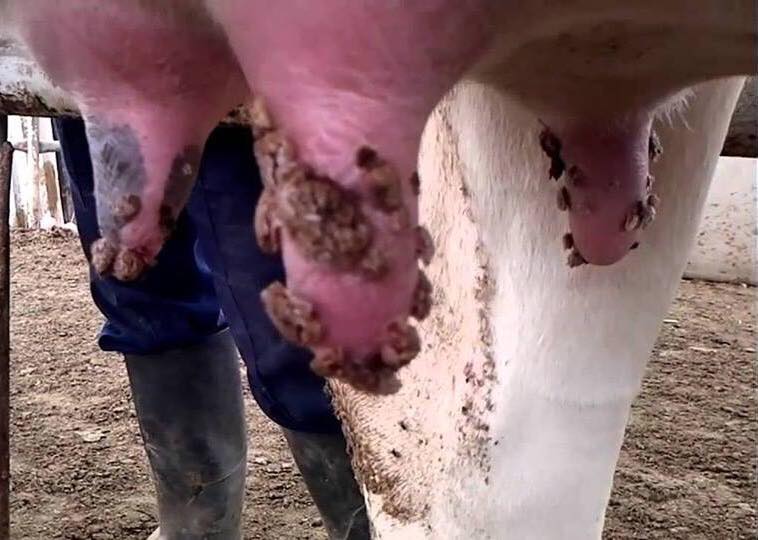
TREATMENT OF TEAT/UDDER WARTS/ Cutaneous Papilloma (Bovine Teat Papillomatosis) IN DAIRY COWS
by-DR RAJESH KUMAR SINGH, JAMSHEDPUR, JHARKHAND,INDIA, 9431309542,rajeshsinghvet@gmail.com
Warts occur quite commonly on cattle in India, especially on young cattle. Warts are contagious and can spread rapidly when cattle are in close contact. They are rarely serious and usually disappear spontaneously. Warts usually occur on the head, neck and shoulders, but can be found anywhere on the animal. They are usually small and cause little trouble. In severe cases, large areas may be involved and animals so affected may not thrive. The warts will bleed if knocked and may become infected or flyblown. Warts may also occur on the udder and vulva of cows, on the penis and prepuce of bulls, and around the anus. Warts reduce the value of hides.
Warts are caused by infection with the contagious bovine papillomavirus. The common wart, Verruca vulgaris, is a specific type of epithelial skin overgrowth, non-malignant in character. Warts are of common occurrence in cattle of all age groups on various parts of the body. The four most common types are squat, pedunculated, and flat and tags. Though warts are little harm and disappear spontaneously over long time, but with recurrence they degrade the value of animal. Surgery and vaccination, or a combination of both, are the most common forms of treatment and prevention. There are no present literatures which reveal the exact drug regimen assuring no recurrence.

Papillomavirus is widely distributed in cattle. Cattle are the main source and natural reservoir of infection by the virus; but, halters, ropes, and instruments can serve as a potential source of infection. Not all animals carrying the virus will have warts. It can be transmitted from the inapparent carrier to the susceptible calf.
How Warts Spread
Warts are caused by a host-specific papilloma virus which enters the skin through abrasions. They usually spread by direct contact between cattle. The virus can be spread by ear-tattooing instruments or through scratches caused by barbed wire. The incubation period, that is, the time a wart takes to develop after infection, is normally three to eight weeks but may be longer. The warts usually remain for up to six months and then disappear without treatment. Some last longer.
TREATMENT
- Treatment is not usually required, as most warts eventually regress spontaneously.
- Surgical removal is possible but may lead to recurrence.
- Removal should only be done on mature growths, since removing warts too soon can stimulate the growth and spread the virus.
- Large pedunculated warts can be removed slowly by tying a ligature around the base. The wart will dry up and fall off within a month.
- Consult a veterinarian for further advice on treatment.
If the treatment of the infected animals be treated sublingually with Thuja drops (Homeopathy preparation) 200C one ml twice daily for 7 days followed by one ml once a day for next 23 days and Thuja ointment applied over the warts twice daily for 30 days. The warts are found to be completely receded within 30 days and no recurrence seen on 3 years observation.
Dairy Cattle Treatment of Teat/Udder Wart/Cutaneous Papilloma (Bovine Teat Papillomatosis) with Auto-Hemotherapy:
This is a treatment that consists of removing blood from Jugular veins of cattle and injecting it back into the muscular tissue. Doing that would increase the amount of macrofags in the body. Macrofages are the front line cells that enter defensive reactions in the body, they constantly circulate throughout all organs with the only aim – to find and remove undesired elements.
“It is a simple and low cost therapeutic resource which is nothing more than drawing blood from a vein and applying it into a muscle. This stimulates the Reticulo-endothelial System and increases fourfold the macrophages in the whole organism.”
This method has been used for over 100 years and nearly disappeared when antibiotics appeared in the 1940’s.
Today due to the use of auto-hemotherapy on a large scale by field veterinarians in India to treat the disease like Cutaneous papillomatosis, there is a popular movement in favour of its formal acceptance.
What is auto-hemotherapy?—
It is a simple technique where by drawing blood from a vein and injecting it into a muscle stimulates an increase of macrophages that are, let us say, the “municipal cleaning company” of the organism.
The macrophages carry out a cleansing of everything. They eliminate bacterias, viruses and cancerous cells which are called neoplasic cells. They do a spring-cleaning, and even eliminate the fibrin, which is clotted blood. The increase in the production of macrophages by the bone marrow occurs because the blood in the muscle works as a foreign body to be rejected by the Reticulo-endothelial System (RES). While there is blood in the muscle, the Reticulo-endothelial System is being activated. The maximum activation only finishes at the end of five days.
The normal rate of macrophages in the blood is 5% (five percent) and with the auto-hemotherapy, we raise this rate to 22% (twenty-two percent) during 5 (five) days. From the 5th (fifth) to the 7th (seventh) day, it starts to drop, because the blood in the muscle is coming to an end. And when it finishes the rate returns to 5% (five percent). This is the reason why the technique dictates the auto-hemotherapy should be repeated every 7 (seven) days.
This is the reason why auto-hemotherapy works. It is a very low cost method, needing only a syringe. It can be done anywhere because it doesn’t depend even on a refrigerator – simply because the blood is drawn in the moment it is applied into the patient, nothing being required to be done to the blood. There is no technique applied to this blood, only a person who knows how to puncture a vein and apply an injection into a muscle with hygiene, and a syringe to draw the blood and apply it into the muscle, nothing else. This results in a very powerful immune stimulant.
Therefore, it is really a method that could be disseminated and used in rural India without any resources, where people can’t afford very expensive immune stimulants
Cutaneous papillomatosis————
Cutaneous papillomatosis is a benign proliferative neoplasm caused by papilloma virus, which usually appear as multiple, sessile or pedunculated, circumscribed greywhite to dark brownish black outgrowth may appear on skin over different body parts. However, neck, eyelids, teats and lower line of abdomen are the most common sites. It is a contagious disease, usually transmitted via direct contact, contaminated food and equipment, flies, castration and injections. Papillomas on teats may cause difficulty in milking and suckling by calf and sometimes, pedunculated papillomas snap-off causing mastitis and teat infections with the clinical signs of various sizes of pedunculated cutaneous warts on various parts of the body with teat involvement, causing pain, bleeding and interference in milking The case is diagnosed as bovine papillomatosis.
Treatment —————–
The Cutaneous papillomatosis involving teat and udder is treated by auto-hemotherapy. Accordingly the animal is administered with its own blood. The venous blood is drawn @ 20 ml from the Jugular vein by using 18G hypodermic needle in a disposable syringe in that 10ml of venous blood is injected subcutaneously in the lateral neck region and 10ml is injected deep intramuscularly in the gluteal region by taking all sterile precautions. The treatment is repeated once in a week for four weeks continuously. After third injection, the papilloma growths shows signs of regression. The animal is under observation for six weeks. By the end of six weeks all the papilloma growths are completely reduced.
Bovine papillomatosis is a contagious disease of cattle occurring as warts on skin and mucosa, caused by Bovine Papilloma Virus types 1 to 10 .The virus infects the basal cells of the epithelium causing the excessive growth, which is characteristic of wart formation. Venugopalan (2000) and O’Conor (2001) have suggested remedial measures for removal of warts such as use of autogenous vaccine, wart enucleation, burning with hot iron or eraser, ligation and surgical removal of wart (excision) with surgical knife, application of salicylic acid ointment, di methtyl sulfoxide ointment and potential caustics. Surgical removal of one or two warts was proposed but surgical intervention and vaccination may increase the size of the residual warts and prolong the course of the disease Wadhwa et al. (1992).
Papilloma viruses are the cause of cutaneous warts in cattle and horses. These viruses have considerable host specificity.In cattle, warts can occur on almost any part of the body and these tumors persist for long periods and are discrete, low, flat and circular in appearance. Surgery and vaccination, or a combination of both, are the most common forms of treatment.
Other Treatment of Papillomatosis:——-
Animals are treated by different therapy.
1. Some are treated by anthiomaline, each ml of anthiomaline contains 60mg of Lithium Antimony thiomaliate.15ml/dose, given by i.m at 48 hours interval for four weeks.
2. treatement by topical application of thuja ointment, thrice a day for four weeks.
3 treatement by oral administration of thuja extract 20gm, thrice a day for four weeks
4. treatement by autohaemotherapy. Accordingly the cow is treated using its own blood.20ml of venous blood is drawn from the Jugular vein using 18G hypodermic needle in a disposable syringe. Each 10ml of it is injected both the sides of the lateral neck region by taking all sterile precautions. The treatment is repeated at weekly interval for four weeks continuously
It is seen that out of the above mentioned line of treatment the treatment by autohaemotherapy gives marvelous result. So we can apply this therapy at field level.
HOMEOPATHY TREATMENT—
Thuja drops 200C one ml twice daily for 7 days followed by one ml once a day for next 23 days and Thuja ointment should be applied over the warts twice daily for 30 days, we get good results at field level.
Prevention
Commercial vaccines are available in developed country (but in India it is yet to come).; and if used as directed, they may help prevent warts in cattle not previously infected. Autogenous vaccines are prepared from chemically treated warts taken from animals in a herd. In fact, the autogenous vaccine is more apt to have the strain or type of papillomavirus causing the wart problem in a herd than some of the commercial vaccines.
Instruments and tack used on infected animals should be disinfected before use on other animals. The infected animal may not have visible warts, but they may still contaminate equipment. Tattoo or tagging pliers can be disinfected between use on calves, with a 2 to 4% solution of formaldehyde. Dilute the liquid formalin 1 to 18 for a 2% solution or 1 to 9 for the 4% solution. Rinse off blood or tissue from the pliers before immersing in the formaldehyde. Maintain two sets of the instruments and alternate them in use thereby providing adequate time in the formaldehyde to inactivate the virus. Rinse them before using and wear examination gloves or rubber household gloves to protect hands from irritation. Tack that has been in contact with infected calves can also be disinfected with formaldehyde.


